Journal of Medical Cases, ISSN 1923-4155 print, 1923-4163 online, Open Access
Article copyright, the authors; Journal compilation copyright, J Med Cases and Elmer Press Inc
Journal website http://www.journalmc.org

Case Report

Volume 6, Number 9, September 2015, pages 433-436


A Surgical Case of Heterotopic Thymic Carcinoma Coexisting With Hepatocellular Carcinoma

Figures

Figure 1.
Figure 1. CT findings over a 7-year period. A small nodule was observed in May 2002 (a), and by June 2006 this had grown to an elliptical shape (b), finally becoming a 4 × 3 cm tumor compressing the right ventricle by February 2009 (c). Arrow: tumor.
Figure 2.
Figure 2. The undifferentiated carcinoma showed sheet-like proliferation without any particular structure or differentiation (a), consisting of small round cells (b).
Figure 3.
Figure 3. The squamous carcinoma showed solid nests of polygonal cells with keratinization (a) and consisted of large irregular nuclei with prominent nucleoli (b).
Figure 4.
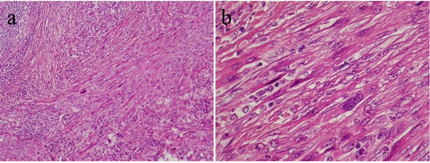
Figure 4. The sarcomatoid carcinoma showed palisade formation and polygonal spindle cells invading the surrounding area (a), and consisted of multinucleated cells (b).