Journal of Medical Cases, ISSN 1923-4155 print, 1923-4163 online, Open Access
Article copyright, the authors; Journal compilation copyright, J Med Cases and Elmer Press Inc
Journal website http://www.journalmc.org

Case Report

Volume 6, Number 11, November 2015, pages 527-533


“Unclassical” Combination of Smell Dysfunction, Altered Abdominal Nociception and Human Hypertension Associated “Classical” Adrenal-Augmentation

Figures

Figure 1.
Figure 1. Arterial-phase enhanced computed tomography. Axial view (left), coronal view (right). Images show a homogenous solid mass with a diameter of about 4 cm, in left hemiabdomen (white arrow). The mass has a well-demarcated margin, and homogeneously low density. Calcification, necrosis or cystic degeneration was not observed. The mass located within the left adrenal gland is also compromising left kidney. The atrophy and loss of cortico-medullary relationship of the left kidney was noteworthy. Asymmetrical renal volume was evident; however, renal obstruction was not.
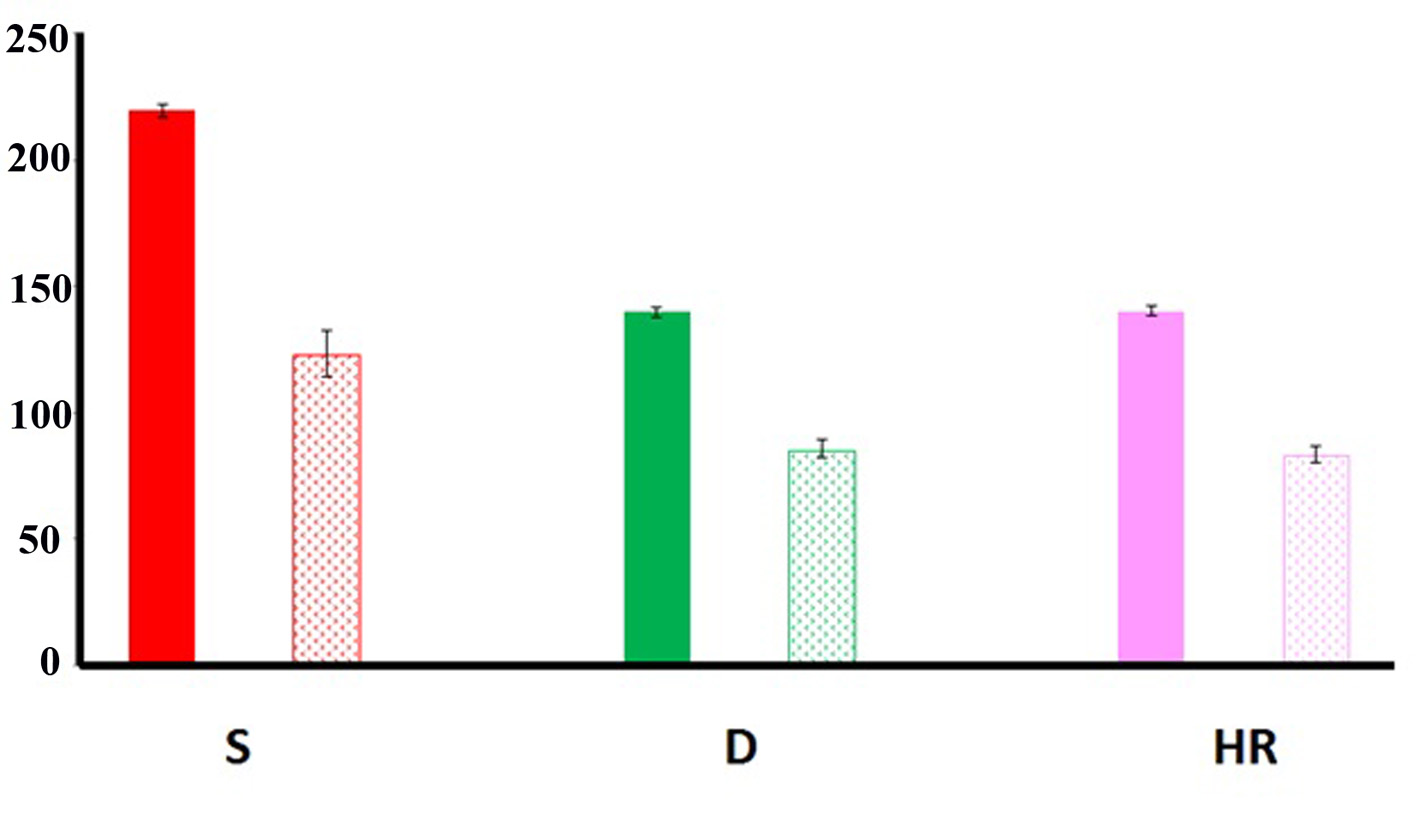
Figure 2.
Figure 2. Autonomic function. Systolic (S), diastolic (D) and heart rate (HR) values obtained before (filled bars) and after (dotted bars) surgical removal of the adrenal adenoma. Note that the values obtained after surgery significantly improved (see text for details). Bars: standard deviation.
Figure 3.
Figure 3. Nociceptive evaluation. The values obtained by applying the visual analog scale (VAS) significantly decreased after mass removal (AS) compared to the values obtained before surgical intervention (BS) (see text for details). Bars: standard deviation.
Figure 4.
Figure 4. Smell function. Scores of the olfactory function measured by the UPSIT before (BS) and after surgery (AS). Note the significant improvement of the smell function found AS (see text for details). Bars: standard deviation.

Tables

Table 1. Summary of the Main Glands and Cells Involved in the Different Types of Multiple Endocrine Neoplasia
 
Target organMEN-IMEN-II (2a)MEN-III (2b)
*The commonalities of such multiple neoplasia. **Thyroid. ***Peripheral nerves.
Pancreas gland+
Pituitary gland+
Parathyroid gland*+++
Chromatin cells*+++
Medullar cells**++
Schwann cells***+


Table 2. Frequency of Tumors Located at the Adrenal Gland
 
LocationType of tumorFrequency
CortexAdenoma+++
Carcinoma++
MedullaPheocromocytoma++
Ganglioneuroma+
Paraganglioma+
Ganglioneuroblastoma+
Mielolipoma+
StromaFibroma+
Hemangioma+
Sarcoma+