Journal of Medical Cases, ISSN 1923-4155 print, 1923-4163 online, Open Access
Article copyright, the authors; Journal compilation copyright, J Med Cases and Elmer Press Inc
Journal website https://www.journalmc.org

Case Report

Volume 14, Number 8, August 2023, pages 293-298


A Case of Radiation-Associated Vertebral Compression Fracture Mimicking Solitary Bone Metastasis of Lung Cancer

Figures

Figure 1.
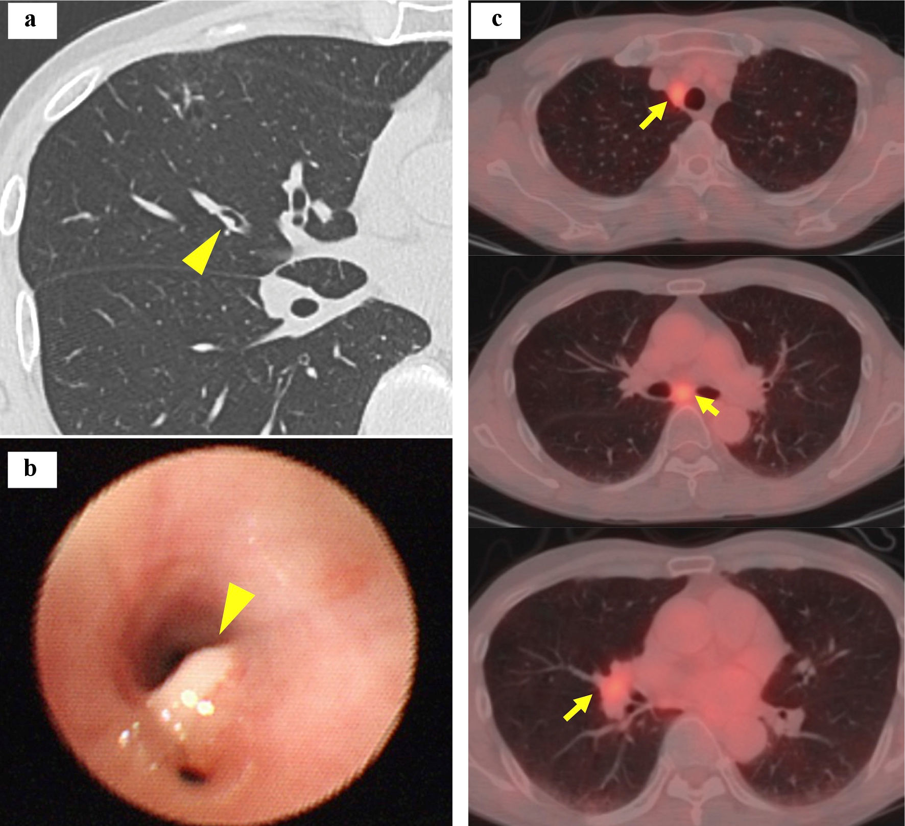
Figure 1. Chest computed tomography (a), a bronchoscopic image of the right B4 (b) and fluorine-18 fluorodeoxyglucose positron emission tomography and computed tomography (c). Yellow arrowhead points to the primary lung cancer (a, b) and yellow arrows point to lymph node metastases (c).
Figure 2.
Figure 2. Histology of bronchoscopial specimen from the tumor in the right B4 (H&E stain). H&E: hematoxylin and eosin.
Figure 3.
Figure 3. Radiation dose distribution and planning design images of chest computed tomography before chemoradiotherapy. Yellow arrowheads point to the seventh thoracic vertebra. The lines represent the irradiation area.
Figure 4.
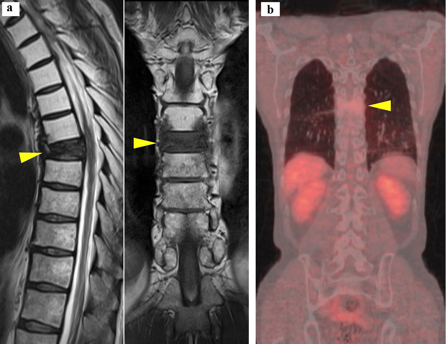
Figure 4. Sagittal (left) and coronal (right) views of magnetic resonance imaging of the spine (a), and coronal view of fused imaging of fluorine-18 fluorodeoxyglucose positron emission tomography and computed tomography (b). Yellow arrowheads point to the fractured seventh thoracic vertebra.
Figure 5.
Figure 5. Histology of the seventh thoracic vertebral specimen (H&E stain). H&E: hematoxylin and eosin.