Journal of Medical Cases, ISSN 1923-4155 print, 1923-4163 online, Open Access
Article copyright, the authors; Journal compilation copyright, J Med Cases and Elmer Press Inc
Journal website https://www.journalmc.org

Case Report

Volume 14, Number 11, November 2023, pages 356-361


Multiple Complications of Crohn’s Disease and the Need for Early and Continuous Multidisciplinary Undertaking

Figures

Figure 1.
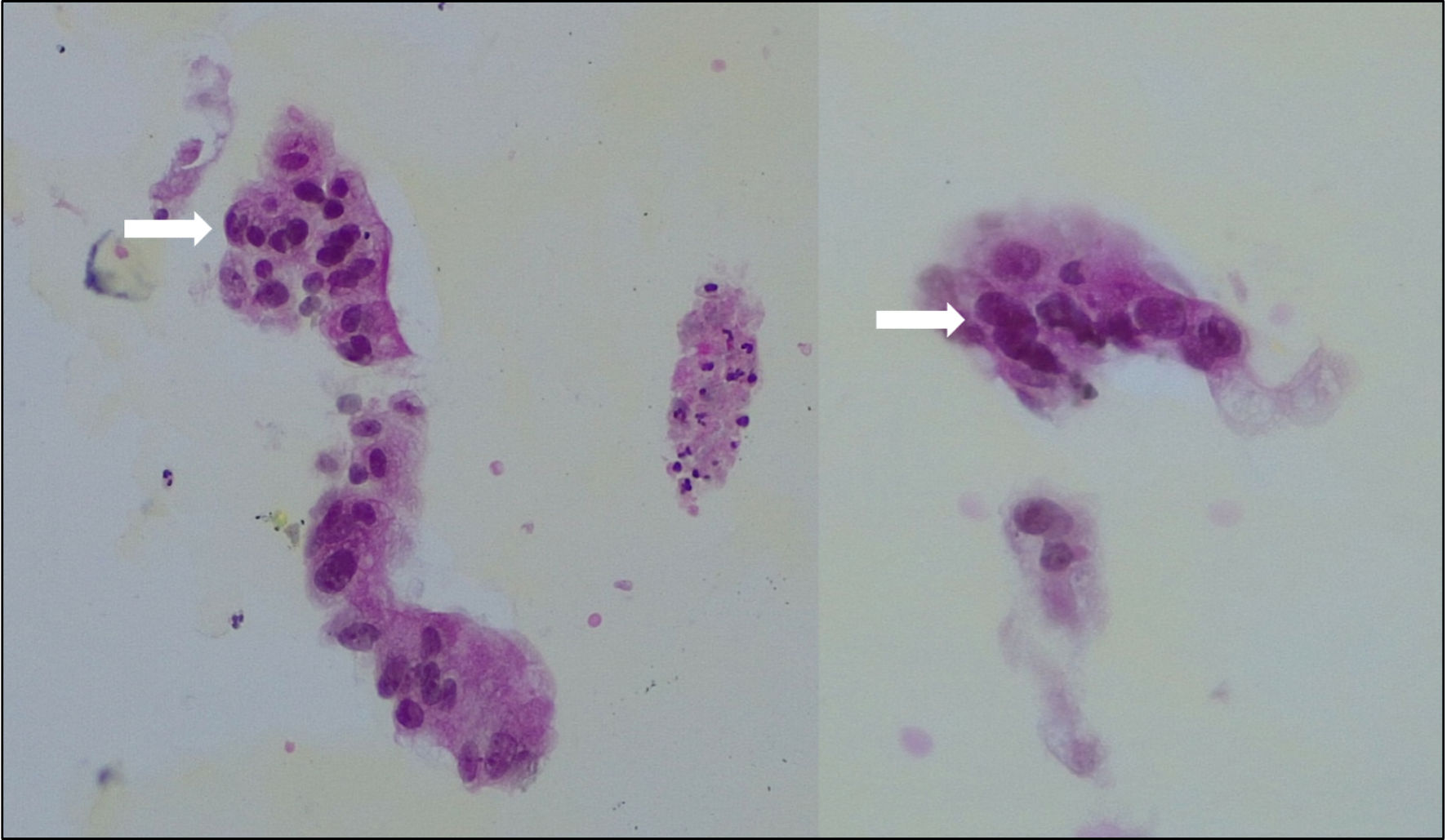
Figure 1. Biopsy of the abdominal mass showing large cylindric cells with important anisonucleosis (arrows), consistent with adenocarcinoma.
Figure 2.
Figure 2. 18F-FDG PET/CT. Sagittal CT view (a) and axial fusion imaging view (b). Voluminous heterogeneous mass along the course of the urachus with heterogeneous FDG uptake. PET/CT: positron emission tomography/computed tomography; FDG: fluorodeoxyglucose; CT: computed tomography.
Figure 3.
Figure 3. Abdominal MRI. T2-weighted sagittal view (a) and T1-weighted axial view after gadolinium injection (b). Heterogeneous enhancement of the mass. MRI: magnetic resonance imaging.
Figure 4.
Figure 4. Abdominal CT scan without contrast, sagittal (a) and axial (b) views. Midline heterogeneous solid lesion (asterisk) overhanging the antero-superior wall of the bladder and extending toward the umbilicus. CT: computed tomography.