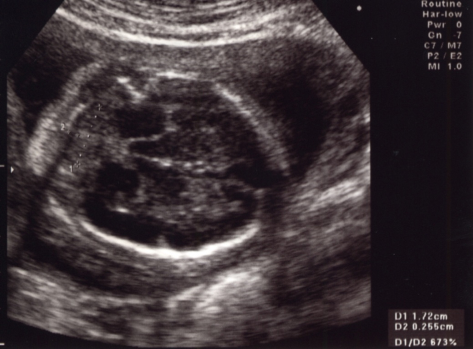
Figure 1. Prenatal ultrasound findings in a fetal thorax at the 25th week of gestation. Horizontal view of the thorax demonstrates a left-side diaphragmatic hernia and mediastinal shift.
| Journal of Medical Cases, ISSN 1923-4155 print, 1923-4163 online, Open Access |
| Article copyright, the authors; Journal compilation copyright, J Med Cases and Elmer Press Inc |
| Journal website http://www.journalmc.org |
Case Report
Volume 4, Number 4, April 2013, pages 215-217
A Case of Trisomy 9 Complicated With Congenital Diaphragmatic Hernia
Figures