Journal of Medical Cases, ISSN 1923-4155 print, 1923-4163 online, Open Access
Article copyright, the authors; Journal compilation copyright, J Med Cases and Elmer Press Inc
Journal website http://www.journalmc.org

Case Report

Volume 4, Number 5, May 2013, pages 292-295


Idiopathic Azygos Vein Aneurysm Mimicking a Mediastinal Mass: Case Report and Review of Literature

Figures

Figure 1.
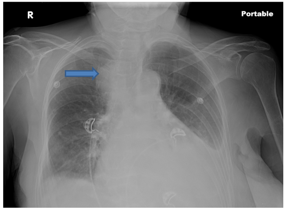
Figure 1. Chest roentgenogram showing a right paratracheal mass with left pleural effusion.
Figure 2.
Figure 2. An axial thoracic contrast-enhanced CT image shows a well-defined 5.5 cm mass (thick arrow) in mediastinum. The mass enhances remarkably and homogeneously in the venous phase, with isodensity to the large vessels in the mediastinum, indicating a vascular origin.
Figure 3.
Figure 3. Computed to tomographic scan of chest (coronal section) showing the azygous arch aneurysm.
Figure 4.
Figure 4. The 3-dimensional CT reconstruction image showing the Azygos aneurysm (arrow).