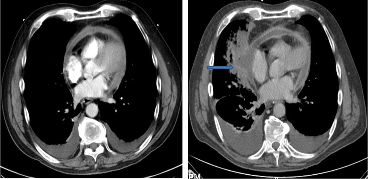
Figure 1. Bilateral pleural effusion with airspace disease involving the right middle and lower lobes.
| Journal of Medical Cases, ISSN 1923-4155 print, 1923-4163 online, Open Access |
| Article copyright, the authors; Journal compilation copyright, J Med Cases and Elmer Press Inc |
| Journal website http://www.journalmc.org |
Case Report
Volume 4, Number 5, May 2013, pages 307-310
A Curious Case of Pericardiohydroptysis
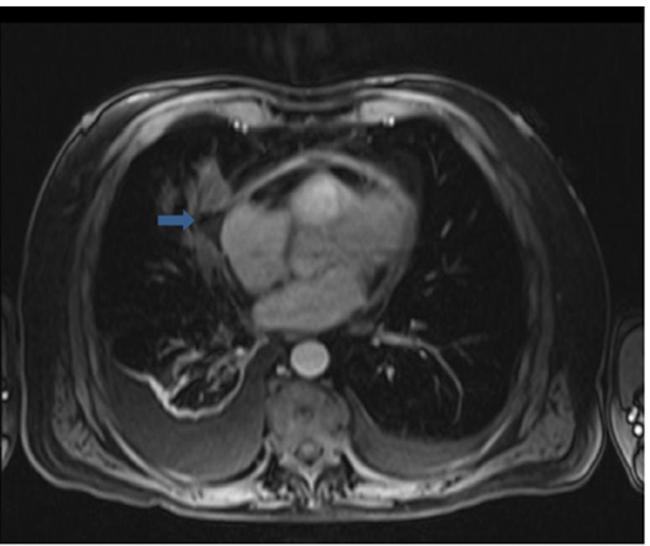
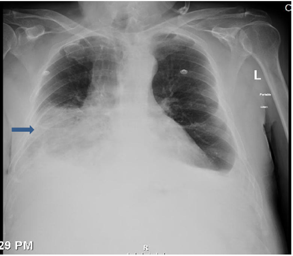
Figures