Journal of Medical Cases, ISSN 1923-4155 print, 1923-4163 online, Open Access
Article copyright, the authors; Journal compilation copyright, J Med Cases and Elmer Press Inc
Journal website http://www.journalmc.org

Case Report

Volume 2, Number 1, February 2011, pages 31-33


Soft Tissue Squamous Cell Carcinoma in Chest Wall with an Incomplete Border Sign

Figures

Figure 1.
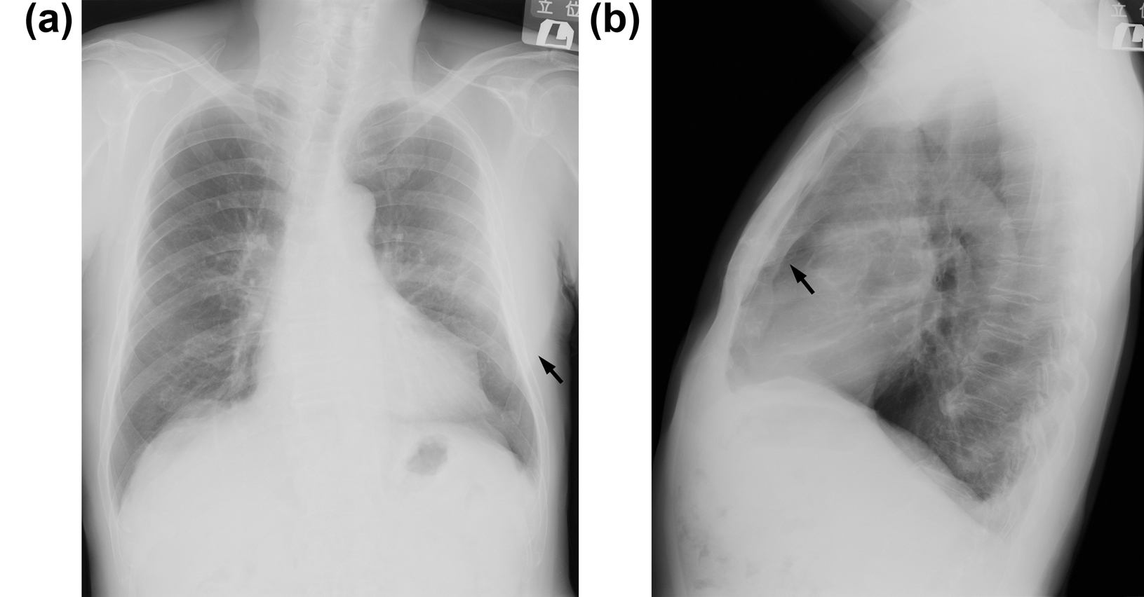
Figure 1. (a) Posteroanterior view of chest radiography revealed a radio-opaque lesion in the left lung field by a sharp, well-defined edge on the left lateral side (arrow). An ill-defined edge on the other side along at least one portion of its contour blends with the chest wall. (b) Oblique view showed an extra pleural mass with a sharp margin (arrow).
Figure 2.
Figure 2. (a) Contrast-enhanced axial CT image showed a mass under the musculus pectoralis major invading the left third rib (arrow). (b) The same level of high-resolution CT (3 mm) revealed focal thickness of the pleura and interstitial reactions (arrow).
Figure 3.
Figure 3. The histological finding obtained from ultrasound-guided needle biopsy confirmed the diagnosis of squamous cell carcinoma (hematoxiline and eosine).