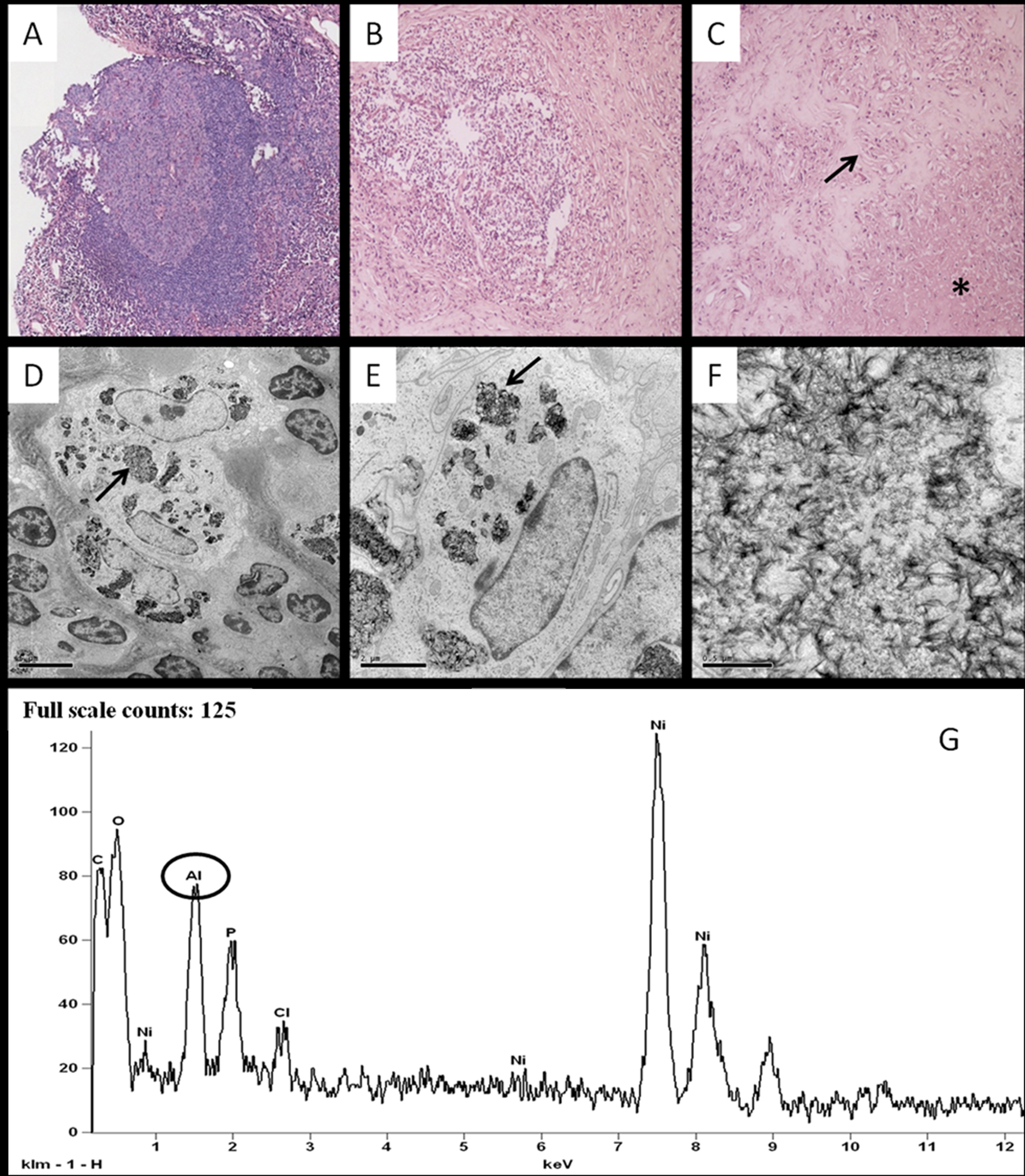
Figure 1. Panel A to C: hematoxylin eosin section of the lesion showing a granulomatous reaction with lymphatic follicle formation (panel A magnification 4 ×), macrophages and lymphoplasmacellular infiltrate (panel B, MAGNIFICATION 10 ×).The lesion is characterized by a necrotic center (asterisk in panel C) surrounded by palisading epithelioid histiocytes (arrow in panel C). PANEL D to F: conventional transmission electron microscopy characterization of the lesion. PANEL D shows a big histiocyte’s granule (arrow) containing fibrillar electron dense material (MAGNIFICATION 3,500 ×). The ultrastructural study demonstrated the presence of needle-shaped crystalline material with variable thickness (PANEL E 10,000 × -arrow-; PANEL F 50,000 ×). EDX microanalysis spectrum (PANEL G) reveals the presence of large amounts of aluminum (circle) and of elements referable to the nickel support (Ni) and to biological compounds (P, CL, C and O).