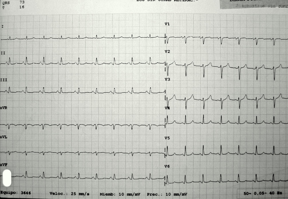
Figure 1. EKG showed Sinus tachycardia, Low-voltage QRS complexes and PR segment depression.
| Journal of Medical Cases, ISSN 1923-4155 print, 1923-4163 online, Open Access |
| Article copyright, the authors; Journal compilation copyright, J Med Cases and Elmer Press Inc |
| Journal website http://www.journalmc.org |
Case Report
Volume 4, Number 7, July 2013, pages 485-487
Cardiac Tamponade Presenting as Abdominal Pain and Being the Initial Manifestation of Malignant Disease: A Case Report
Figures