Journal of Medical Cases, ISSN 1923-4155 print, 1923-4163 online, Open Access
Article copyright, the authors; Journal compilation copyright, J Med Cases and Elmer Press Inc
Journal website http://www.journalmc.org

Case Report

Volume 4, Number 6, June 2013, pages 402-406


Pneumocystis jirovecii and Cytomegalovirus Co-Infection in AIDS Patients

Figures

Figure 1.
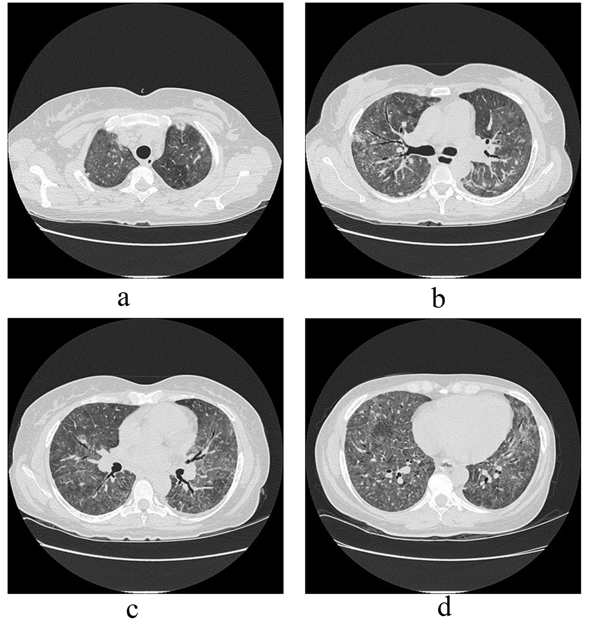
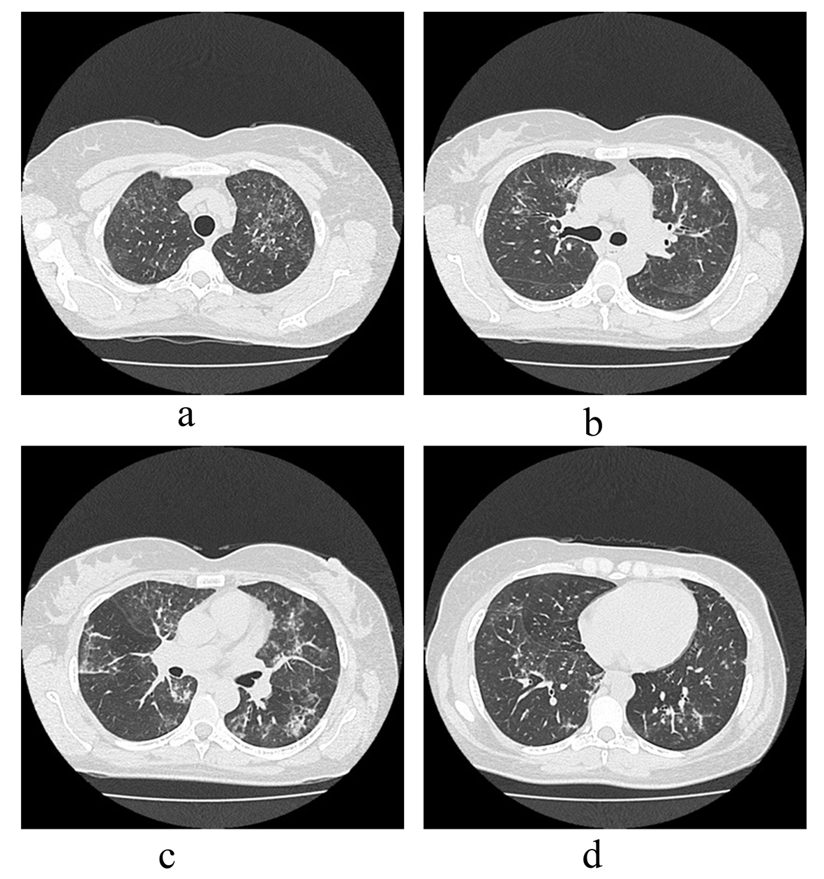
Figure 1. (a-d). Ground-glass opacities (GGO) distributed scarcely in the upper to lower lobes in chest-CT three months before hospitalization.
Figure 2.
Figure 2. (a-d). During the hospitalization, GGO spread in all lobes.
Figure 3.
Figure 3. Lung condensation with foamy and pink exudate (P. jiroveci) filled alveoli (H&E - 200 ×). The alveolar wall shows inflammation, with lymphoid infiltrates and hyaline membranes.
Figure 4.
Figure 4. Violaceous to dark red intranuclear CMV inclusion with foamy exudate fills alveoli associated with Pneumocystis jirovecci (H&E - 100 ×).
Figure 5.
Figure 5. Clusters of Pneumocystis jirovecci (Methenamine silver stain - 100 ×).
Figure 6.
Figure 6. Strong nuclear staining for CMV may be seen.