Journal of Medical Cases, ISSN 1923-4155 print, 1923-4163 online, Open Access
Article copyright, the authors; Journal compilation copyright, J Med Cases and Elmer Press Inc
Journal website http://www.journalmc.org

Case Report

Volume 4, Number 9, September 2013, pages 584-587


Propofol Infusion Syndrome: ACase Report

Figures

Figure 1.
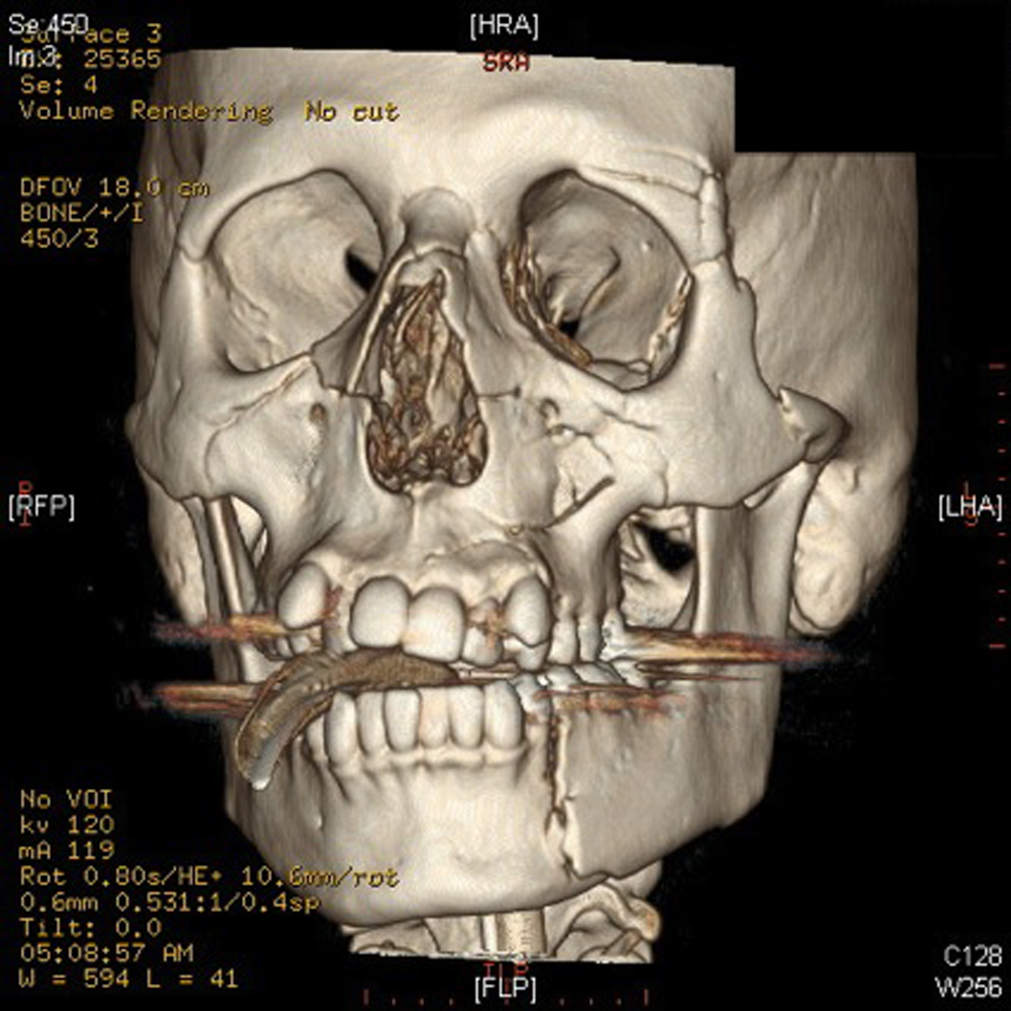
Figure 1. Depicted above is a CT reconstruction of the patient’s facial injuries. These include fractures of the bilateral medial and lateral pterygoid plates, bilateral nasal bones, and left zygoma. Comminuted fractures of the medial and lateral walls of both maxillary sinuses are visualized. There are fractures of the medial and lateral walls of the left orbit, which is comminuted in the lateral wall. There are also bilateral orbital floor fractures, which involve both orbital rims and infra-orbital foramina. There are also several fracture lines extending from the left maxillary sinus into the left maxilla. There is also a fracture through the left mandibular body.
Figure 2.
Figure 2. These 2 electrocardiograms were taken on ICU day 1 and day 4. Notable are new ST elevations in leads V1-V3 in a Brugada-like pattern often seen in patients with propofol-infusion syndrome.

Tables

Table 1. Worsening Rhabdomyolysis, Kidney Injury, Hyperkalemia and Metabolic Acidosis are Evident Throughout the Patients Stay all Indicative of Worsening PRIS
 
WBCCrKTrop ICKpHaHCO3
Day 110.81012.97.3224
Day 27.0893.37.3926
Day 312.01003.37.3528
Day 46.9923.20.2189737.3727
Day 57.5853.40.42524287.3626
Day 618.31316.50.69677437.2015
Day 732.42387.12.918630077.2010


Table 2. Common Risk Factors and Typical Manifestations of PRIS
 
Risk FactorsManifestations
Propofol administration for > 48hrs in doses > 67mcg/kg/min or 5mg/hrRhabdomyolysis
Male sexRenal Failure
Age < 19 years oldHyperkalemia
Head traumaLactic acidosis
Airway infectionHypertriglyceridemia
Increased catecholamine and glucocorticoid levelsBradycardia and Cardiovascular collapse