Journal of Medical Cases, ISSN 1923-4155 print, 1923-4163 online, Open Access
Article copyright, the authors; Journal compilation copyright, J Med Cases and Elmer Press Inc
Journal website http://www.journalmc.org

Case Report

Volume 4, Number 6, June 2013, pages 426-428


A Great Imitator: Sarcoidosis

Figures

Figure 1.
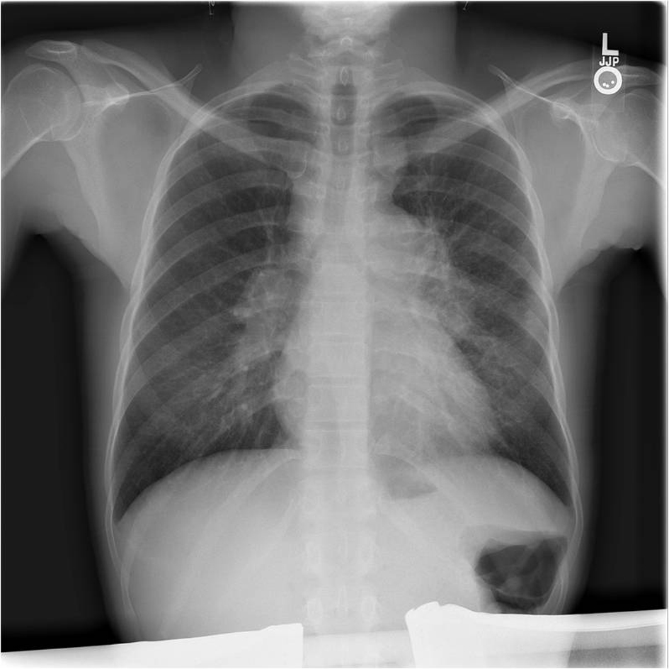
Figure 1. Chest radiograph illustrating mediastinal and bilateral hilar adenopathy.
Figure 2.
Figure 2. Computed tomography of the chest. (a). Axial image through the chest in lung windows illustrating pulmonary nodules (arrows) abutting the right major fissure. (b). Sagittal image through the chest in bone windows shows innumerable sclerotic (white) lesions throughout the sternum and spine.
Figure 3.
Figure 3. Histopathology from sternal biopsy (hematoxylin and eosin stain, 40 ×). A high magnification view shows one of numerous intersitial noncaseating granulomas. The granulomas are well formed and composed of tightly clustered histiocytes. Similar findings were obtained from the transbronchial biopsy.