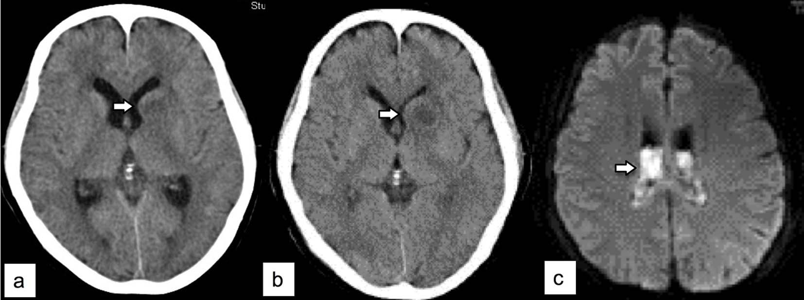
Figure 1. (a) Plain brain computed tomography reveals a low-density lesion (arrow) in the left lenticular nucleus and caudate nucleus, and the left lateral ventricle is compressed. (b) Brain contrast-enhanced CT shows enlarged low-density lesion (arrow) with slight ring enhancement. (c) Brain contrast-enhanced magnetic resonance imaging shows high-intensity areas (arrow) on diffusion-weighted images in bilateral lateral ventricles, indicating rupture of the abscess into the ventricles.