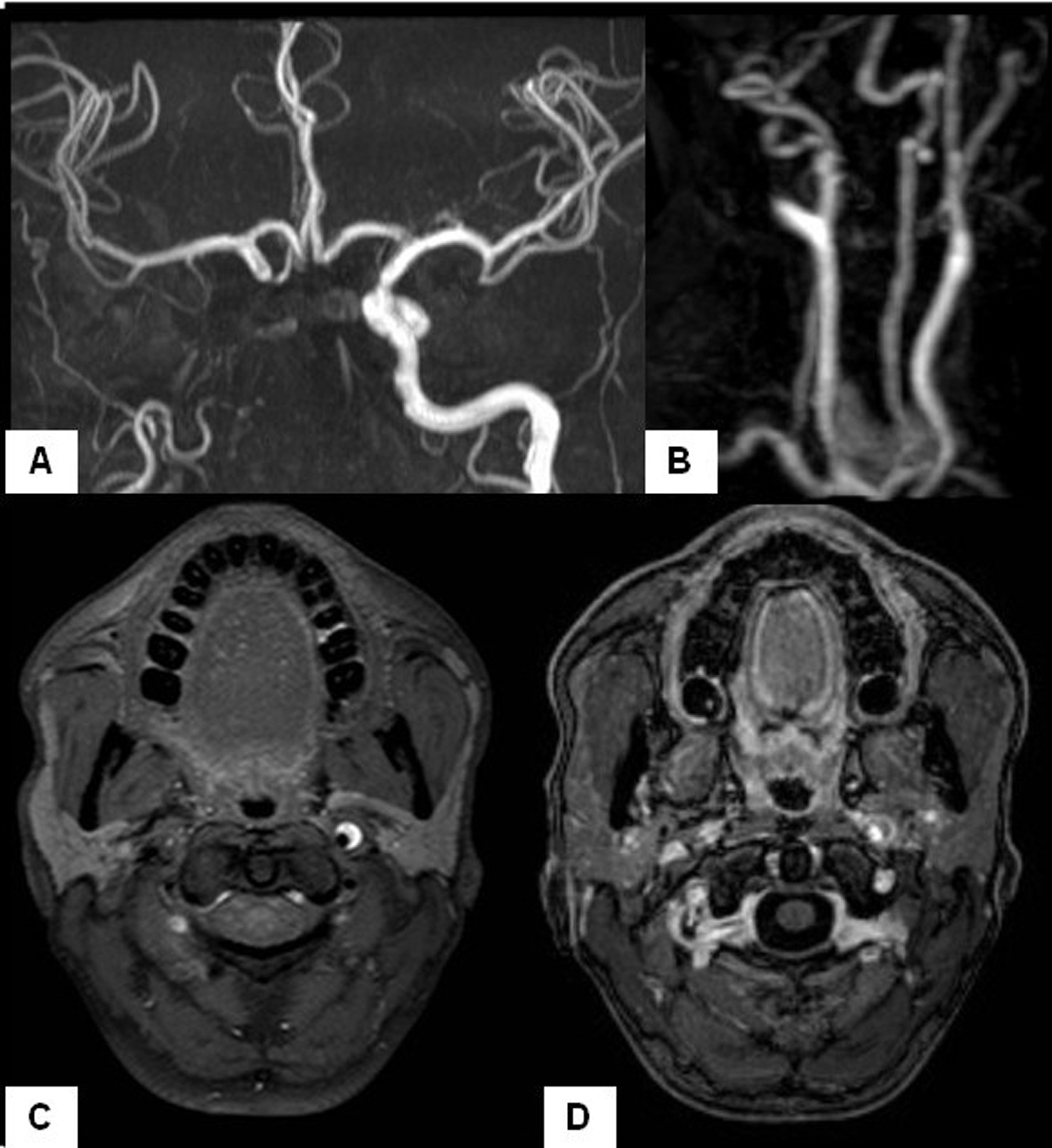
Figure 1. MR-angiography of intracranial (A) and cervical vessels (B) shows the absence of the ICA except for a short proximal segment of sharpened morphology. (C) Cranial MRI shows the typical half-moon hyperintense signal on the left ICA wall before its entrance into the petrous canal, consistent with dissection (T1-weighted, fat saturation sequences). Notice the absence of representation of the right carotid artery at the same level. (D) After gadolinium administration no decrease in the lumen of the left ICA was observed, also indicating arterial dissection.