Journal of Medical Cases, ISSN 1923-4155 print, 1923-4163 online, Open Access
Article copyright, the authors; Journal compilation copyright, J Med Cases and Elmer Press Inc
Journal website http://www.journalmc.org

Case Report

Volume 4, Number 9, September 2013, pages 623-626


Computed Tomography Findings of a Case of Megacolon Associated with Caudal Regression Syndrome

Figures

Figure 1.
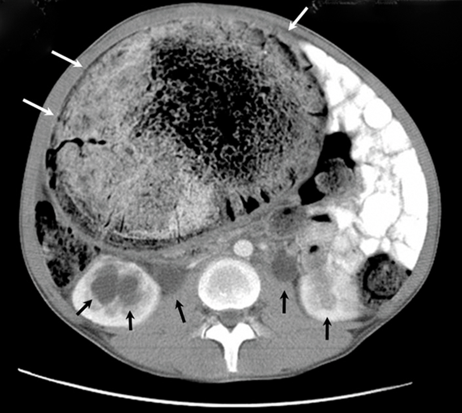
Figure 1. An axial CT scan shows the huge, dilated, non-haustrated sigmoid colon (white arrows), bilateral dilated urinary calices, and ureters (black arrows). The thickness of the bowel wall is normal, and there is no contrast enhancement. The compressed and displaced small bowel on the left side of the abdomen can also be seen.
Figure 2.
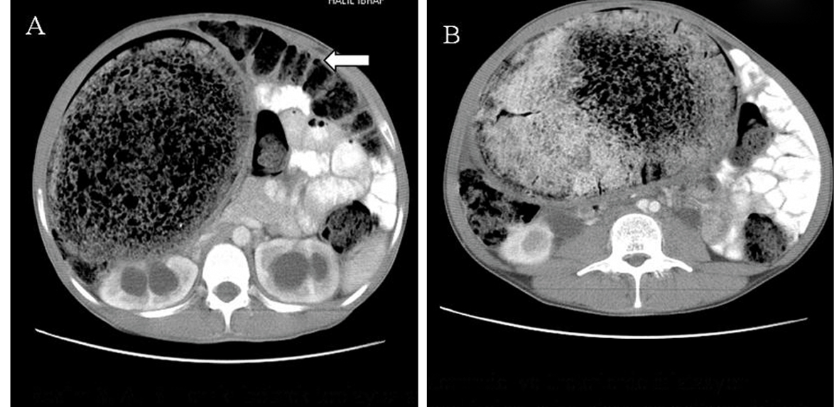
Figure 2. A, B. Axial CT scans show the huge, dilated, non-haustrated sigmoid colon. The loops of the large bowel are also dilated and filled with feces. They are not filled with oral contrast material, and their normal haustration pattern is preserved. The compressed and displaced small bowel on the left side of the abdomen is also shown.
Figure 3.
Figure 3. An axial CT scan shows the anteriorly displaced bladder with irregular wall thickening (black arrows) along with the absence of inferior sacrococcygeal vertebrae (white arrows).
Figure 4.
Figure 4. An axial CT scan shows the absence of inferior sacral vertebrae (black arrows). Abdominal distension secondary to the huge, dilated bowel which fills the whole pelvis is also shown.