Journal of Medical Cases, ISSN 1923-4155 print, 1923-4163 online, Open Access
Article copyright, the authors; Journal compilation copyright, J Med Cases and Elmer Press Inc
Journal website http://www.journalmc.org

Case Report

Volume 4, Number 9, September 2013, pages 644-647


Endobronchial Metastases of Renal Cell Carcinoma: A Complex Multidisciplinary Treatment

Figures

Figure 1.
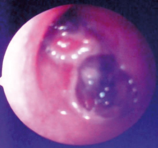
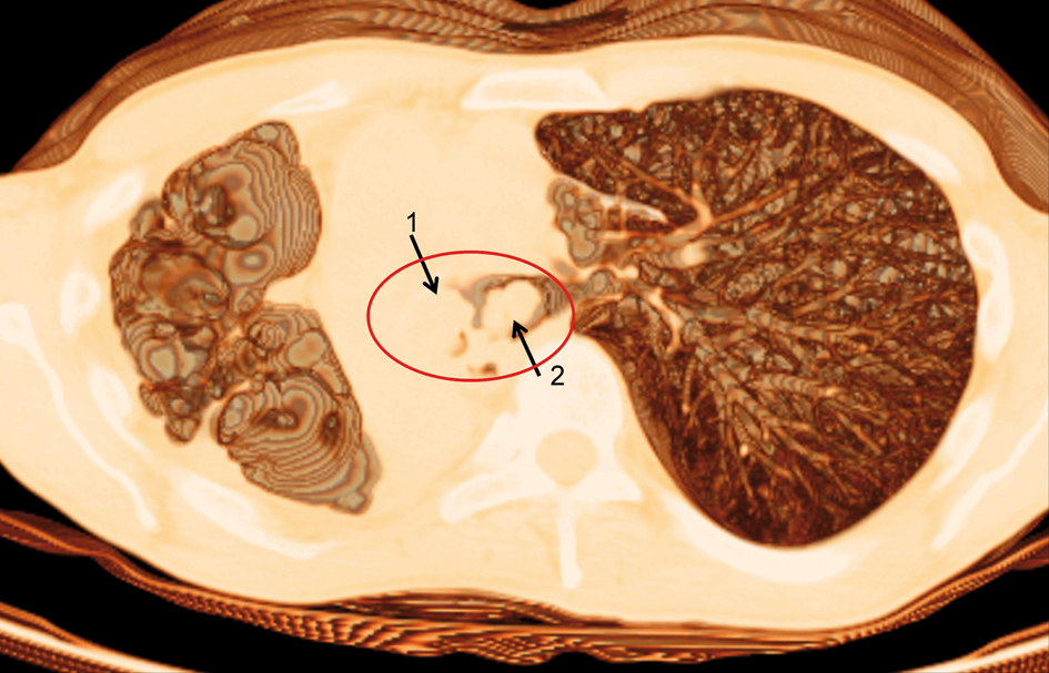
Figure 1. Computed tomography scan of the chest showing the tracheal carina (circle) with complete obstruction of the left main bronchus (arrow 1) and stenosis of the right main bronchus (arrow 2).
Figure 2.
Figure 2. Endoscopic view of the main bronchi before laser resection.
Figure 3.
Figure 3. Right intercostal bronchial artery (arrow 1) that supply the neoplasm (arrow 2). Superselective bronchial artery embolization of the right main bronchus with injection of microbeads (250 - 300 micron) and the positioning of four platinum spirals compatible with magnetic resonance imaging (arrow 3).
Figure 4.
Figure 4. Upper left bronchial artery before (arrow 1) and after (arrow 2) selective embolization of with microbeads (250 - 300 micron).
Figure 5.
Figure 5. Flexible bronchoscopy showing the complete reopening of the right main bronchus (arrow).