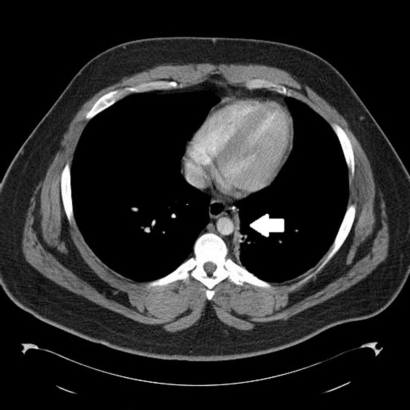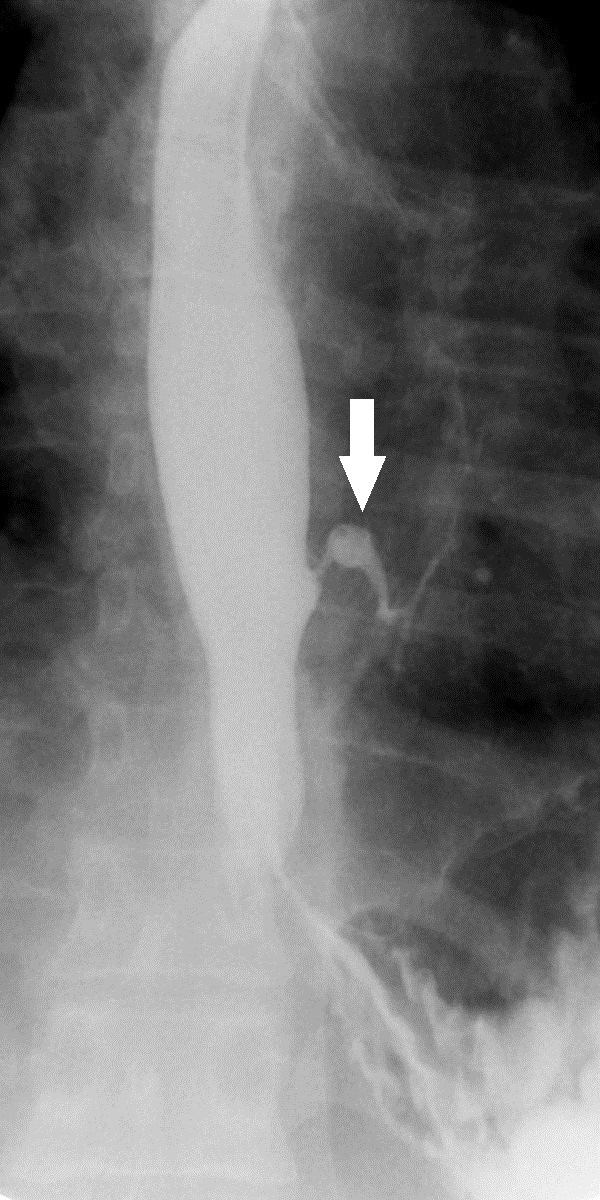
Figure 1. Spot fluoroscopic image from a single contrast esophagram demonstrates a fistulous tract between the lateral esophageal wall and the left lower lobe bronchus.
| Journal of Medical Cases, ISSN 1923-4155 print, 1923-4163 online, Open Access |
| Article copyright, the authors; Journal compilation copyright, J Med Cases and Elmer Press Inc |
| Journal website http://www.journalmc.org |
Case Report
Volume 2, Number 2, April 2011, pages 64-66
A Rare Benign Bronchoesophageal Fistula Presenting in Adulthood: A Case Report
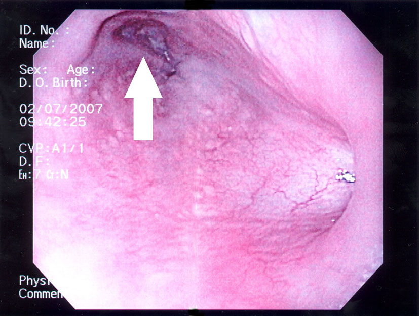
Figures