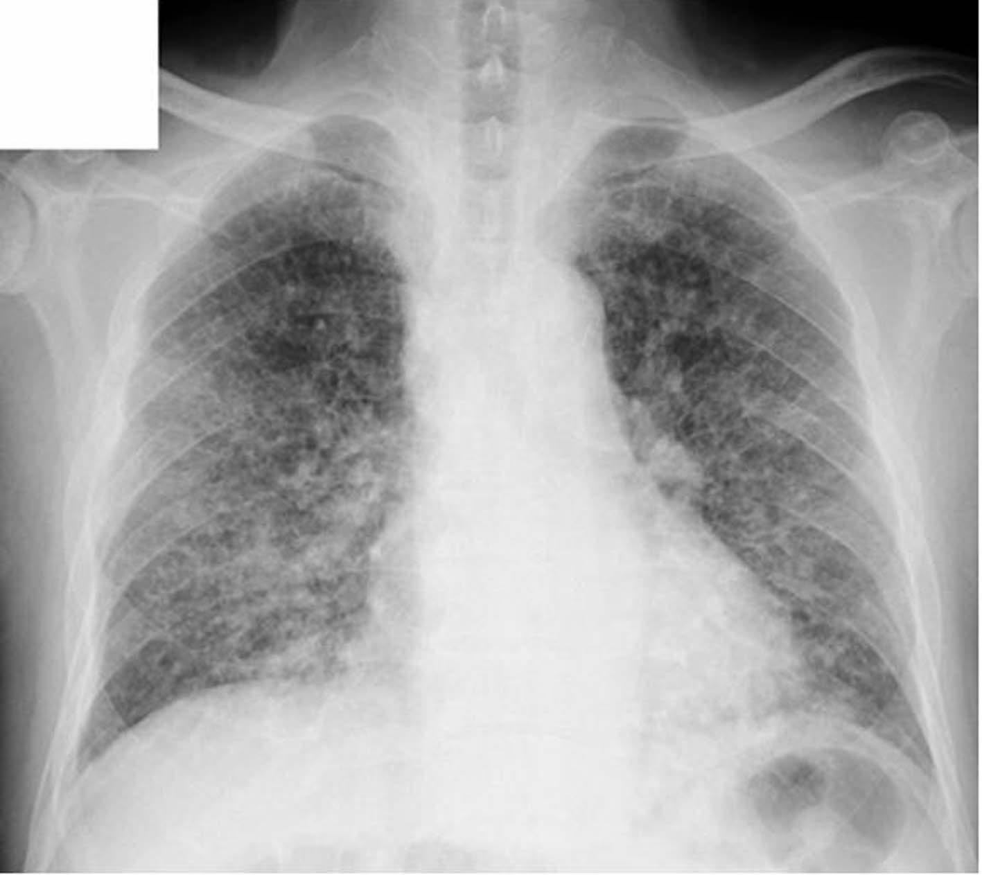
Figure 1. The chest X-ray in the initial diagnosis.
| Journal of Medical Cases, ISSN 1923-4155 print, 1923-4163 online, Open Access |
| Article copyright, the authors; Journal compilation copyright, J Med Cases and Elmer Press Inc |
| Journal website http://www.journalmc.org |
Case Report
Volume 2, Number 2, April 2011, pages 81-86
A Case of Prostate Carcinoma Discovered With Pulmonary Lymphangitis Carcinomatosa
Figures
Table
| CBC, complete blood count; WBC, white blood cell; RBC, red blood cell; BBC, blood biochemistry; AST, aspartate aminotransferase; ALT, alanine aminotransferase; LDH, lactate dehydrogenase; γ-GTP, γ-glutamyltranspeptidase; CPK, creatine phosphate kinase; TP, total protein; UA, uric acid; BUN, blood urea nitrogen; Cre, creatinine; Glu, glucose; HbA1c, hemoglobin A1c; CRP, C reactive protein; CEA, carcinoembryonic antigen; sLI-2R, serum interleukin II receptor; PR3, proteinase-3; ANCA, antineutrophil cytoplasmic antibody; MPO, myeloperoxidase; ABG, arterial blood gas analysis; BE, base excess; SaO2, Oxygen saturation. | |||||
| CBC | Normal range | Normal Range | |||
| WBC | 12600/µl | (3900 – 9300) | Cre | 0.92 mg/dl | |
| RBC | 511 x 104/µl | Na | 141 mEq/L | ||
| hemoglobin | 14.5 g/dl | K | 4.0 mEq/L | ||
| hematocrit | 43.6% | Cl | 99 mEq/L | (70 - 109) | |
| platelet | 19 x 104/µl | Glu | 221 mg/dl | (4.3 - 5.8) | |
| Hemogram | HbA1c | 8.5% | (< 0.3) | ||
| neutrophil | 76% | CRP | 28.4 mg/dl | ||
| lymphocyte | 16.8% | CEA | 0.5 ng/ml | ||
| monocyte | 6.3% | Ca 19-9 | 11.1 U/ml | (124 - 466) | |
| eosinophil | 0.3% | sIL2R | 564 U/ml | ||
| basophil | 0.6% | KL-6 | 257 U/ml | ||
| BBC | SP-D | 17.2 ng/ml | |||
| T-Bil | 1.9 mg/dl | (0.2 - 1.0) | PR3-ANCA | < 10 EU | |
| AST | 95 U/L | (5 - 40) | MPO-ANCA | < 10 EU | |
| ALT | 48 U/L | (5 - 45) | ABG | ||
| LDH | 1293 U/L | (120 - 240) | PH | 7.464 | |
| γ-GTP | 187 U/L | (≤ 85) | PCO2 | 31 mm Hg | (32 - 46) |
| CPK | 133 U/L | PO2 | 79 mm Hg | ||
| TP | 6.8 g/dl | HCO3- | 22.0 mmol/L | ||
| UA | 4.5 mg/dl | BE | -0.6 mmol/L | ||
| BUN | 25.9 mg/dl | (8.0 - 20.0) | SaO2 | 96.9% | |