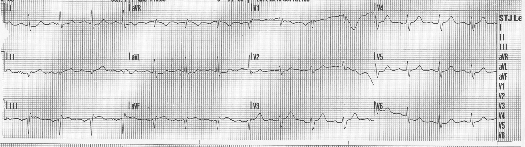
Figure 1. 12-lead EKG done en route to the hospital showing ST elevations and q waves in inferior leads.
| Journal of Medical Cases, ISSN 1923-4155 print, 1923-4163 online, Open Access |
| Article copyright, the authors; Journal compilation copyright, J Med Cases and Elmer Press Inc |
| Journal website http://www.journalmc.org |
Case Report
Volume 5, Number 2, February 2014, pages 73-75
Acute Pulmonary Embolism Masquerading as Acute Inferior Myocardial Infarction
Figures