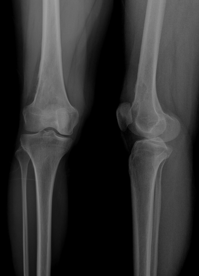
Figure 1. Film obtained 2 years ago during the patient’s first admission.
| Journal of Medical Cases, ISSN 1923-4155 print, 1923-4163 online, Open Access |
| Article copyright, the authors; Journal compilation copyright, J Med Cases and Elmer Press Inc |
| Journal website http://www.journalmc.org |
Case Report
Volume 5, Number 6, June 2014, pages 315-325
Bizarre Parosteal Osteochondromatous Proliferation of Long Bones: Two New Cases and Literature Study
Figures
Tables
| No. of papers | Study | Year | No. of patients | Sex/age | Trauma | Symptom | Involved bone | Localization |
|---|---|---|---|---|---|---|---|---|
| S&P: swelling and pain; PM: proximal metaphysic; DM: distal metaphysic; PEM: proximal epyphisiometaphysis; DEP: distal epyphisiometaphysis. | ||||||||
| 1 | Gruber [16] | 2008 | 1 | M/16 | No | S&P | Ulna | DM |
| 2 | Bhalla [18] | 2012 | 1 | M/10 | Yes | S&P | Femur | DM |
| 3 | Kershen [21] | 2012 | 1 | F/37 | No | S&P | Tibia | Diaphysis |
| 4 | Rybak [10] | 2007 | 2 | F/16 | No | S | Ulna | DM |
| M/36 | Radius | DM | ||||||
| 5 | Abramovici [3] | 2002 | 2 | F/12 | No | S&P | Femur | DM |
| M/27 | Yes | S&P | Tibia | PM | ||||
| 6 | Joseph [9] | 2011 | 1 | F/22 | Yes | S | Tibia | PM |
| 7 | Meneses [2] | 1993 | 17 | - | - | - | Humerus (1) Ulna (6) Radius (3) Femur (3) Fibula (2) Tibia (2) | Not defined |
| 8 | Berber [6] | 2011 | 5 | Not defined | Not defined | No | Radius | PM DM PM DM |
| Ulna | DM | |||||||
| Tibia | PM | |||||||
| Femur (2) | DM | |||||||
| 9 | Cooper [11] | 1993 | 1 | M/37 | No | S | Radius | Diaphysis |
| 10 | Helliwell [17] | 2001 | 1 | M/15 | Yes | S | Radius | Diaphysis |
| 11 | Bush [4] | 2007 | 1 | F/51 | Yes | S&P | Humerus | PEM |
| 12 | Boudova [19] | 1999 | 1 | M/52 | Yes | P&S | Femur | PM |
| 13 | Vlychou [5] | 2008 | 1 | F/30 | No | S&P | Clavicle | DM |
| 14 | Ly [20] | 2004 | 1 | M/18 | Yes | S&P | Humerus | Diaphysis |
| 15 | Hefferman [14] | 2008 | 1 | F/41 | No | S | Ulna | DM |
| 16 | Nora [1] | 1983 | 2 | M/24 F/21 | Not defined | S | Humerus Radius | DM DM |
| 17 | Author cases | New case | 1 | M/19 | Yes | P | Femur | DEP |
| New case | 1 | M/21 | Yes | S&P | Femur | DM | ||
| 2006[12] | 1 | F/27 | No | S&P | Femur | DM | ||
| 18 | Choi [15] | 2001 | 1 | F/18 | No | S | Fibula | DM |
| No. of papers | X-ray | CT | MRI | Size of tumor (cm) | C. Dest | M. Inv | Stage |
|---|---|---|---|---|---|---|---|
| C. Dest: cortical destruction; M. Inv: medullary invasion. | |||||||
| 1 | Cortical-based calcified and osseous masses | Intensely calcified and ossified masses | Marrow involvement: No Soft-tissue extension: No | Not defined | No | No | II |
| 2 | Broad-based osseous protuberance masses, Cortical reactive sclerosis | Exostotic appearing lesion | Marrow involvement: No Soft-tissue extension: No | 3 | Yes | No | III |
| 3 | Cortical-based calcified and osseous masses | Not defined | Marrow involvement: No Soft-tissue extension: No | 5 | No | No | II |
| 4 | 1. Pedunculated mass | Pedunculated mass of mature ossification with distinct medullary and cortical components | Not defined | 3 | Yes | Yes | IV |
| 2. Cortical-based calcified and osseous masses | Not defined | Not defined | Not defined | Yes | Not defined | II | |
| 5 | 1. Cortical involvement and destruction as well as focal calcification | Cortical destruction, Cortical reactive sclerosis | Marrow involvement: No Soft-tissue extension: No | 4 | Yes | No | III |
| 2. Soft-tissue density mass | Marrow involvement: No Soft-tissue extension: No | 2 cm | No | No | I | ||
| 6 | Well-defined ossified parosteal lesion | Not defined | Not defined | 4 cm | Yes | Not defined | III |
| 7 | Not defined | Not defined | Not defined | Not defined | Not defined | Not defined | No |
| 8 | Not defined | Not defined | Not defined | Not defined | Not defined | Not defined | II |
| 9 | Mushroom-shaped calcified mass, thickening cortex | Not defined | Not defined | 2 | Yes | No | III |
| 10 | Mushroom-shaped calcification mass | Cortical involvement and destruction | Marrow involvement: Yes Soft-tissue extension: Yes | 4 cm | Yes | Yes | IV |
| 11 | A surface based, radiodense lesion | No intramedullary extension and no cortical disruption | Marrow involvement: No Soft-tissue extension: No | 6 | No | No | II |
| 12 | Egg-shaped calcification | No intramedullary extension and no cortical disruption | Not defined | 5 | No | No | I |
| 13 | Ill-defined, radiodense mass | Not defined | Marrow involvement: No Soft-tissue extension: No | Not defined | Yes | No | II |
| 14 | Cortical-based calcified | Calcified soft-tissue mass | Not defined | Not defined | No | No | I |
| 15 | Well-defined ossified lesion | Cortical involvement and destruction | Marrow involvement: No Soft-tissue extension: No | 2 | Yes | No | III |
| 16 | Not defined | Not defined | Not defined | Not defined | Not defined | Not defined | No |
| 17 | 1. Well-defined ossified parosteal lesion, thickening cortex | Not defined | Marrow involvement: No Soft-tissue extension: No | 10 | Yes | No | III |
| 2. Normal | Cortical involvement and destruction | Marrow involvement: Yes Soft-tissue extension: Yes | 3 | Yes | Yes | IV | |
| 3. Ossified parosteal lesion | Cortical involvement and destruction | Marrow involvement: Yes Soft-tissue extension: No | 10 | Yes | Yes | IV | |
| 18 | Cortical-based calcified mass | Not defined | Not defined | 5 | Not defined | No | No |
| No. of papers | Diagnosis | Treatment | Time and No. of recurrence | Recurrence treatment | Complication | Follow-up (months) | Result |
|---|---|---|---|---|---|---|---|
| HE: histopathologicexamination; LAT: local adjuvant therapy; ND: not defined; NR: no recurrence; IE: intralesional curetage. | |||||||
| 1 | HE | Excision | 10 months | Second IE | No | 26 | NR |
| 2 | HE | Excision | ND | Popliteal aneurysm | ND | ND | |
| 3 | HE | Excision | ND | No | ND | ND | |
| 4 | HE | Not defined | 3 years | Excision | No | ND | ND |
| HE | Excision | No | No | No | ND | ND | |
| 5 | HE | Not defined | No | No | No | 12 | NR |
| HE | Not defined | No | No | No | 60 | NR | |
| 6 | HE | M. Resection | No | No | No | 24 | NR |
| 7 | ND | Not defined | ND | ND | ND | ND | ND |
| 8 | HE | Shark-bite | ND | ND | ND | ND | NR |
| Shave exc. | |||||||
| M. Resection | |||||||
| M. Resection | |||||||
| M. Resection | |||||||
| 9 | HE | W. Resection | No | No | No | 24 | NR |
| 10 | HE | W. Resection | No | No | No | 24 | NR |
| 11 | HE | M. Resection | No | No | AVN | 42 | NR |
| 12 | HE | Excision | Four time | Wide excision | No | 18 | NR |
| 13 | HE | Excision | No | No | No | 48 | NR |
| 14 | CT | No surgery | No surgery | No surgery | No surgery | No surgery | No surgery |
| 15 | HE | M. Resection | No | No | No | 12 | NR |
| 16 | HE | Not defined | ND | ND | ND | ND | ND |
| 17 | HE | M. Resection | No | No | No | 90 | NR |
| Curettage LAT | No | No | No | 10 | NR | ||
| Curettage LAT | No | No | No | 12 | NR | ||
| 18 | HE | Excision | ND | No | Fibrosarcoma | ND | ND |