Journal of Medical Cases, ISSN 1923-4155 print, 1923-4163 online, Open Access
Article copyright, the authors; Journal compilation copyright, J Med Cases and Elmer Press Inc
Journal website http://www.journalmc.org

Case Report

Volume 5, Number 3, March 2014, pages 147-151


Pigmented Villonodular Synovitis for Combined Therapy

Figures

Figure 1.
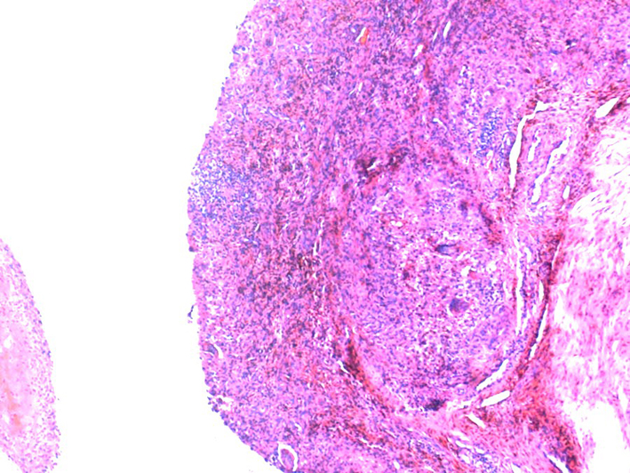
Figure 1. Pigmented villonodular synovitis. Synovia with dense cellular infiltration by mononuclear and giant cells and hemosiderin (brownish areas) deposition. Hematoxylin&eosin × 50. Hemosiderin often forms after bleeding (hemorrhage). When blood leaves a ruptured blood vessel, the red blood cells die and the hemoglobin of the cell is released into the extracellular space. Macrophages engulf (phagocytose) the hemoglobin to degrade it, producing hemosiderin.
Figure 2.
Figure 2. Pigmented villonodular synovitis. Maximal knee flexion after radical synovectomy (left) and combined (surgery + RT) therapy (right: returned to normal).
Figure 3.
Figure 3. Pigmented villonodular synovitis. US longitudinal suprapatellar view study. The synovitis residue after radical synovectomy is seen on the left figure. The rest of synovitis decreased after radiotherapy (right figure).