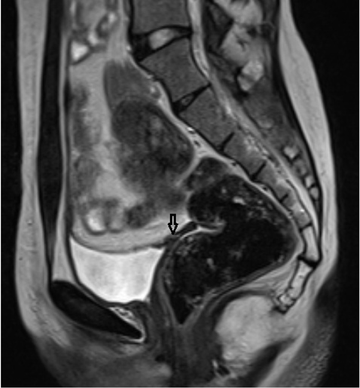
Figure 1. Rudimentary uterus in the retrovesical area in sagittal T2 TSE sequence (arrow).
| Journal of Medical Cases, ISSN 1923-4155 print, 1923-4163 online, Open Access |
| Article copyright, the authors; Journal compilation copyright, J Med Cases and Elmer Press Inc |
| Journal website http://www.journalmc.org |
Case Report
Volume 5, Number 3, March 2014, pages 182-185
Magnetic Resonance Imaging and Clinical Features in Mayer-Rokitansky-Kuster-Hauser Syndrome
Figures