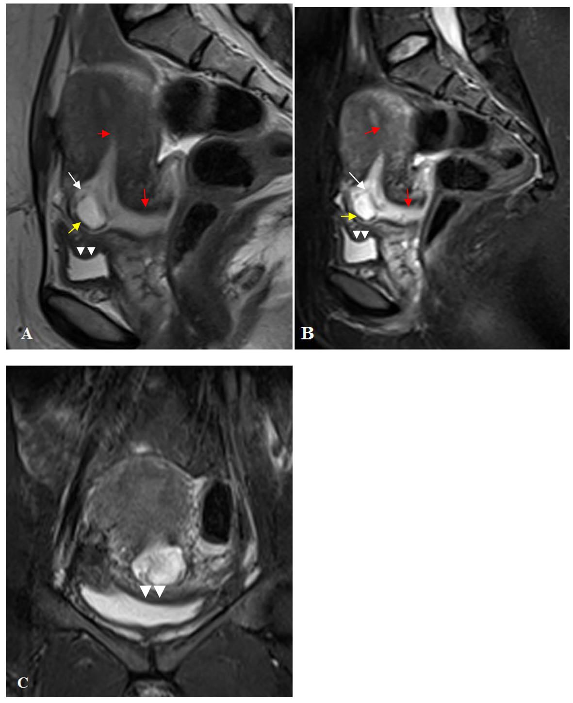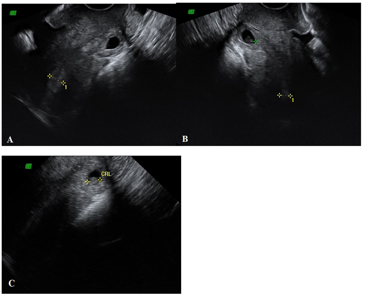
Figure 1. (A, B, C) At the level of the cervical canal there was an ectopic gestational sac including crown-rump length measuring 7.6 mm corresponding to an estimated gestational age of 6 weeks and 5 days.
| Journal of Medical Cases, ISSN 1923-4155 print, 1923-4163 online, Open Access |
| Article copyright, the authors; Journal compilation copyright, J Med Cases and Elmer Press Inc |
| Journal website http://www.journalmc.org |
Case Report
Volume 5, Number 6, June 2014, pages 329-333
Magnetic Resonance Imaging in Addition to Ultrasound in the Diagnosis of Cesarean Scar Ectopic Pregnancy
Figures