Journal of Medical Cases, ISSN 1923-4155 print, 1923-4163 online, Open Access
Article copyright, the authors; Journal compilation copyright, J Med Cases and Elmer Press Inc
Journal website http://www.journalmc.org

Case Report

Volume 5, Number 9, September 2014, pages 505-508


Occult Vertebral Osteomyelitis, a Diagnostic Conundrum

Figures

Figure 1.
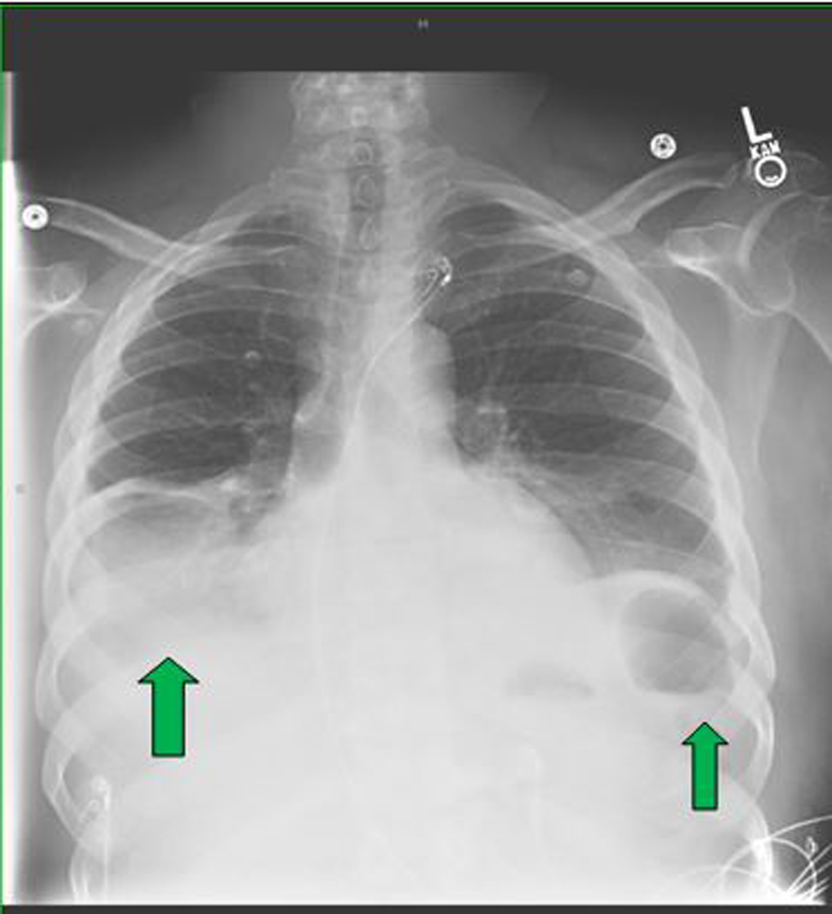
Figure 1. The initial chest X-ray revealed bilateral pleural effusion, markedly greater on the right than the left side (green arrows).
Figure 2.
Figure 2. The CT scan revealed asymmetric pleural effusion more on right pleural space than the left (blue arrows).
Figure 3.
Figure 3. The WBC scan showed increased tracer activity in the left foot suggesting active inflammation (blue circle).
Figure 4.
Figure 4. The whole body WBC scan did not reveal any tracer activity in the body.
Figure 5.
Figure 5. The emergent MRI scan showed T7-8 discitis and osteomyelitis associated with epidural phlegmon resulting in cord compression and edema (red arrows).