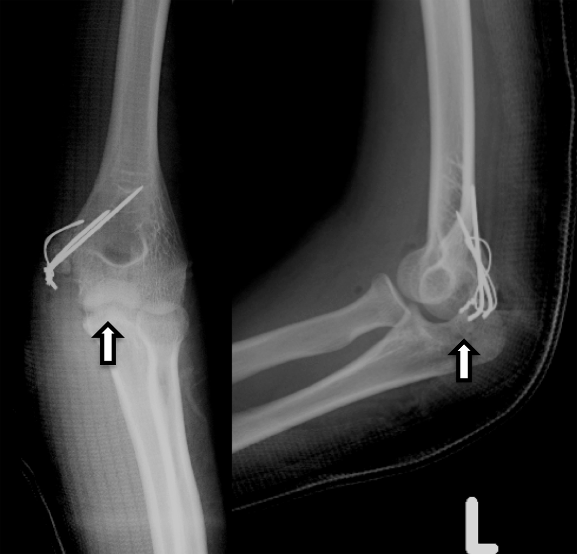
Figure 1. Radiograph obtained soon after the patient’s visit to the general physician (Watson-Jones type 3). Careful retrospective evaluation showed a free interposed bone fragment within the elbow (arrow).
| Journal of Medical Cases, ISSN 1923-4155 print, 1923-4163 online, Open Access |
| Article copyright, the authors; Journal compilation copyright, J Med Cases and Elmer Press Inc |
| Journal website http://www.journalmc.org |
Case Report
Volume 5, Number 7, July 2014, pages 401-403
Interposed Free Bone Fragment in Dislocated Medial Epicondyle Fracture of the Elbow: A Case Report
Figures