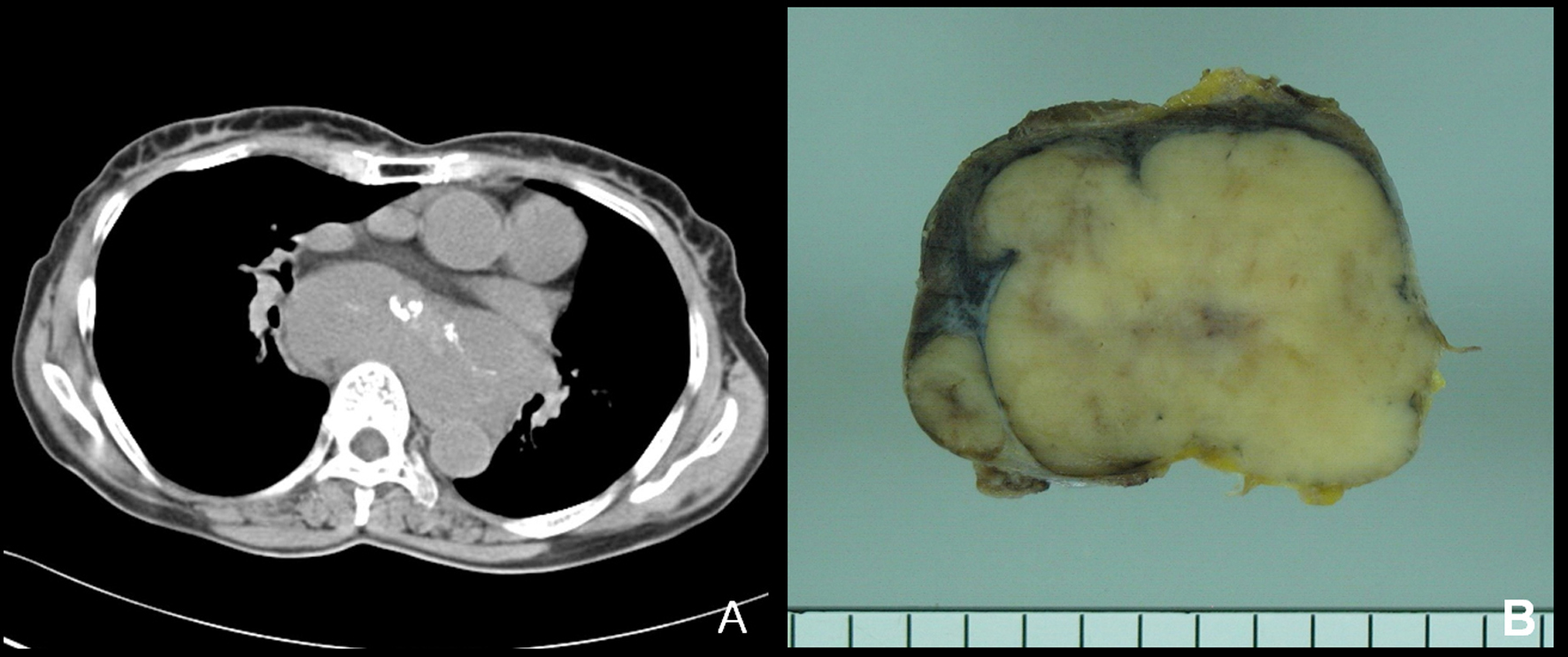
Figure 1. (A) In computed tomography, a huge lobulated solid mass is seen in the posterior mediastinum. (B) Grossly, the resected tumor is solid, vaguely nodular and tan to grayish cut surface. Adjacent enlarged lymph nodes are included.
| Journal of Medical Cases, ISSN 1923-4155 print, 1923-4163 online, Open Access |
| Article copyright, the authors; Journal compilation copyright, J Med Cases and Elmer Press Inc |
| Journal website http://www.journalmc.org |
Case Report
Volume 2, Number 4, August 2011, pages 143-146
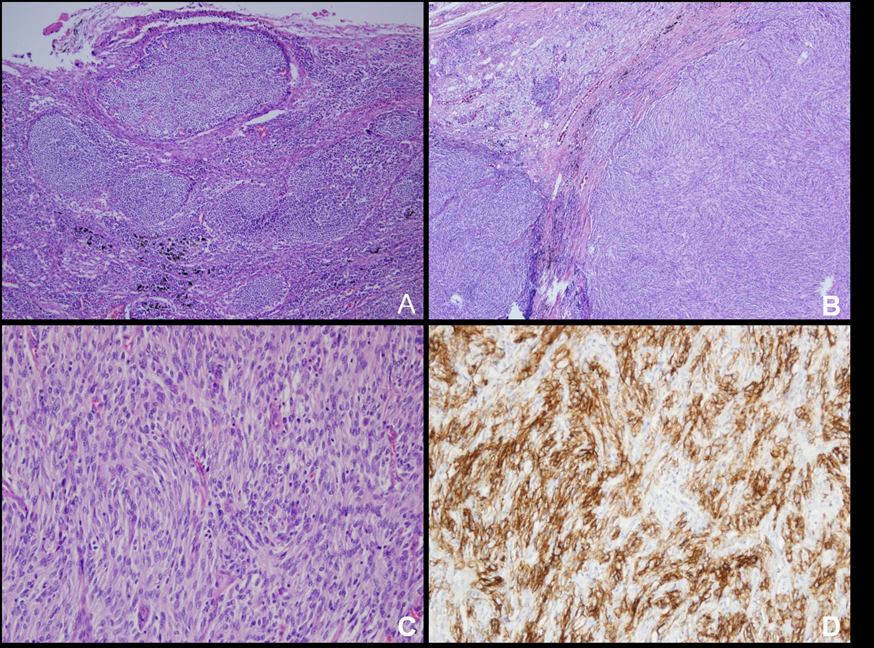
Follicular Dendritic Cell Sarcoma Arising in Hyaline-Vascular Castleman Disease of Mediastinum: A Case Report
Figures