Journal of Medical Cases, ISSN 1923-4155 print, 1923-4163 online, Open Access
Article copyright, the authors; Journal compilation copyright, J Med Cases and Elmer Press Inc
Journal website http://www.journalmc.org

Case Report

Volume 7, Number 7, July 2016, pages 274-281


Supratentorial Extraventricular Anaplastic Ependymoma Presented With Headache in Pregnancy: Case Report and Review of the Literature

Figures

Figure 1.
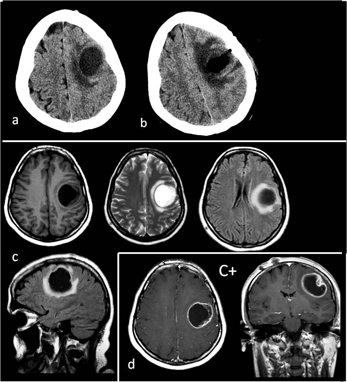
Figure 1. (a) Preoperative tomography to first diagnosis. (b) Postoperative tomography after the first operation. (c) Preoperative T1, T2, flair axial MR images and flair sagittal MR image to first diagnosis. (d) T1 axial and coronal MRI with gadolinium enhancement (C+) to first diagnosis. Tumor appearance cystic, intra-cortical and near cortical surface.
Figure 2.
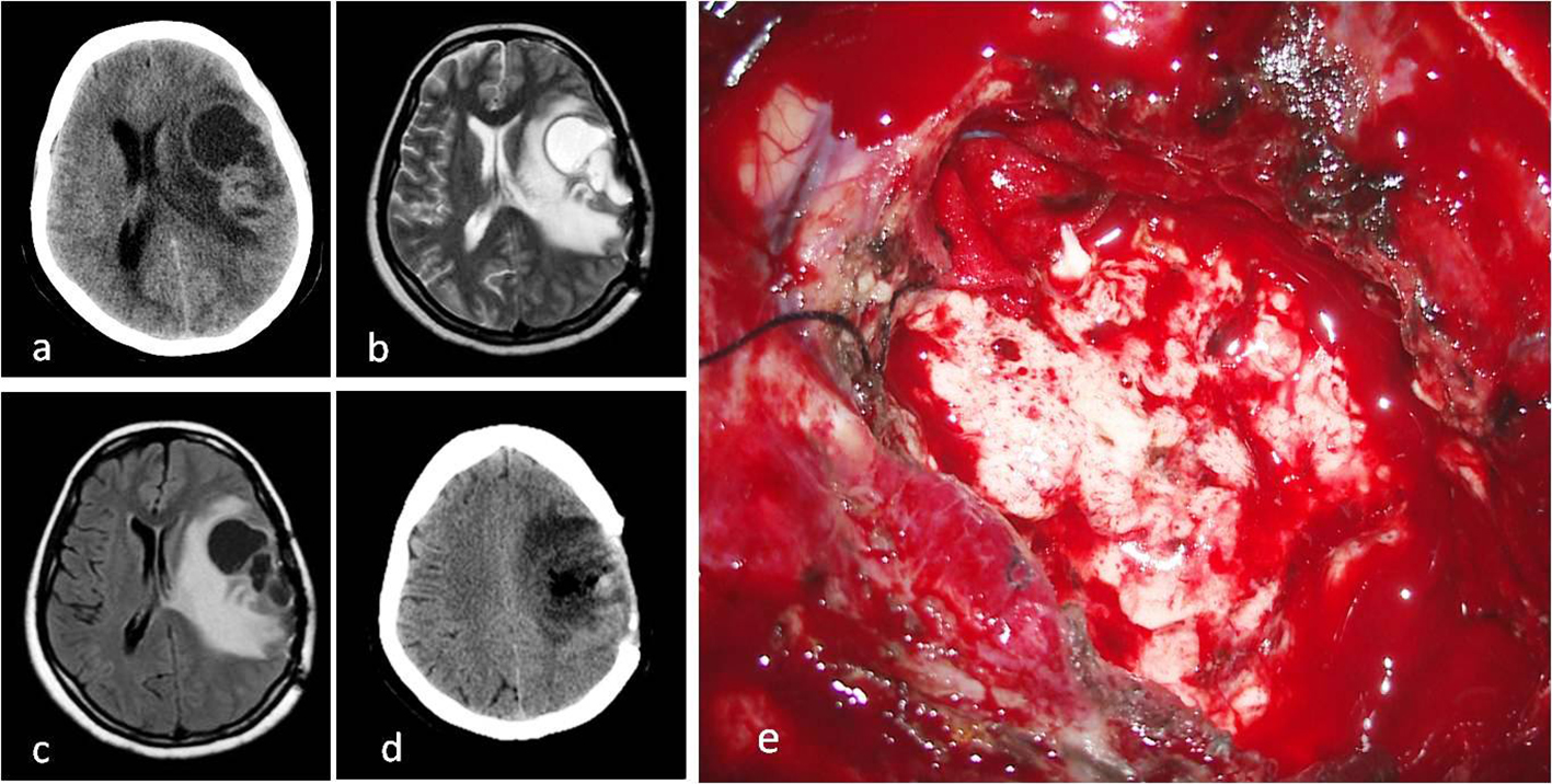
Figure 2. (a) Cystic mass on CT image before second operation. (b) T2 axial MRI. (c) FLAIR axial MRI. (d) CT image after second operation. (e) Intraoperative macroscopic appearance of tumor with surgery microscope.
Figure 3.
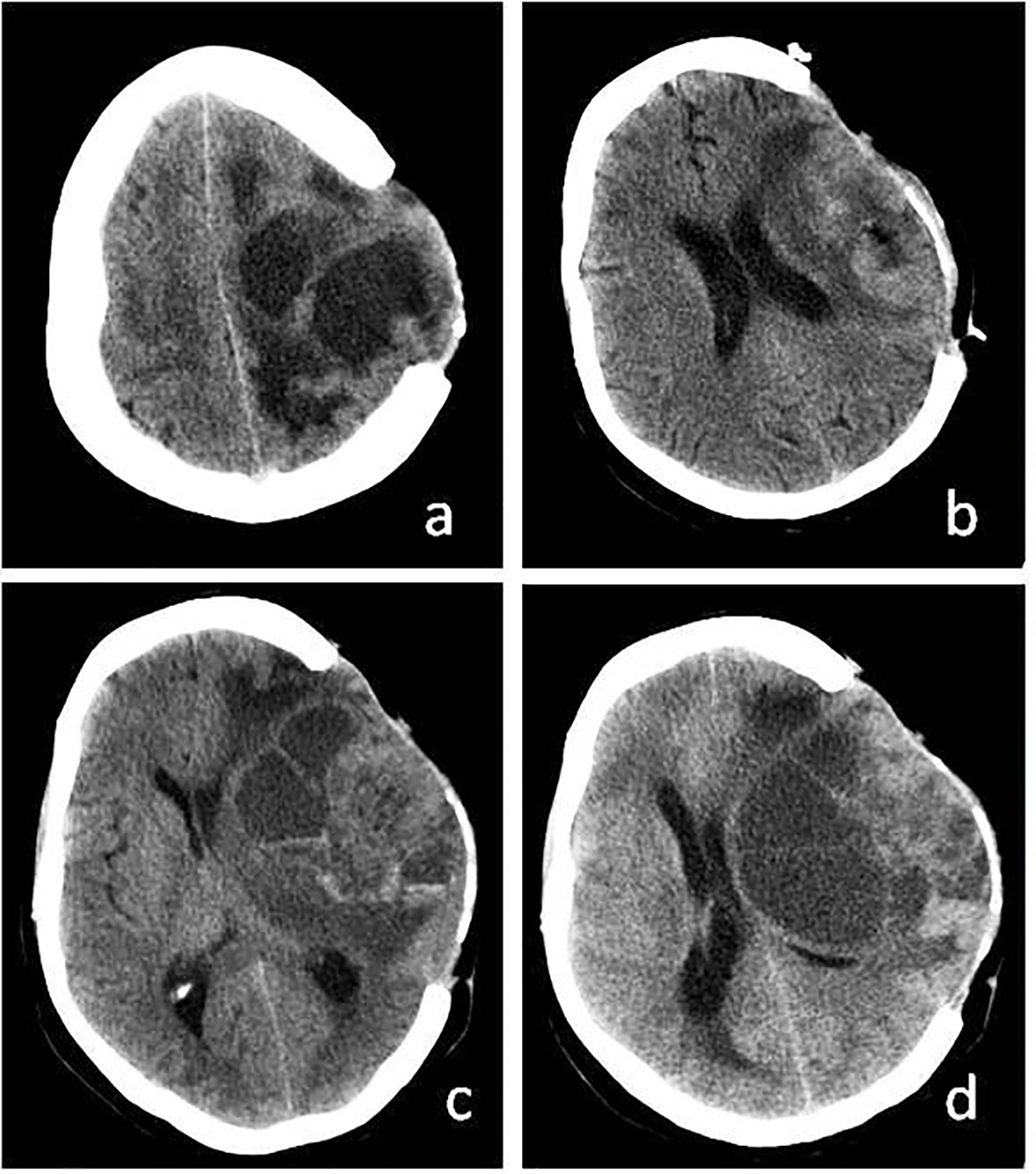
Figure 3. (a) Recurrence of cystic mass on CT before third operation. (b) More calvarial decompression and tumor resection after third operation. (c) Again recurrence of cystic mass. (d) Recurrence of cystic mass before exitus.
Figure 4.
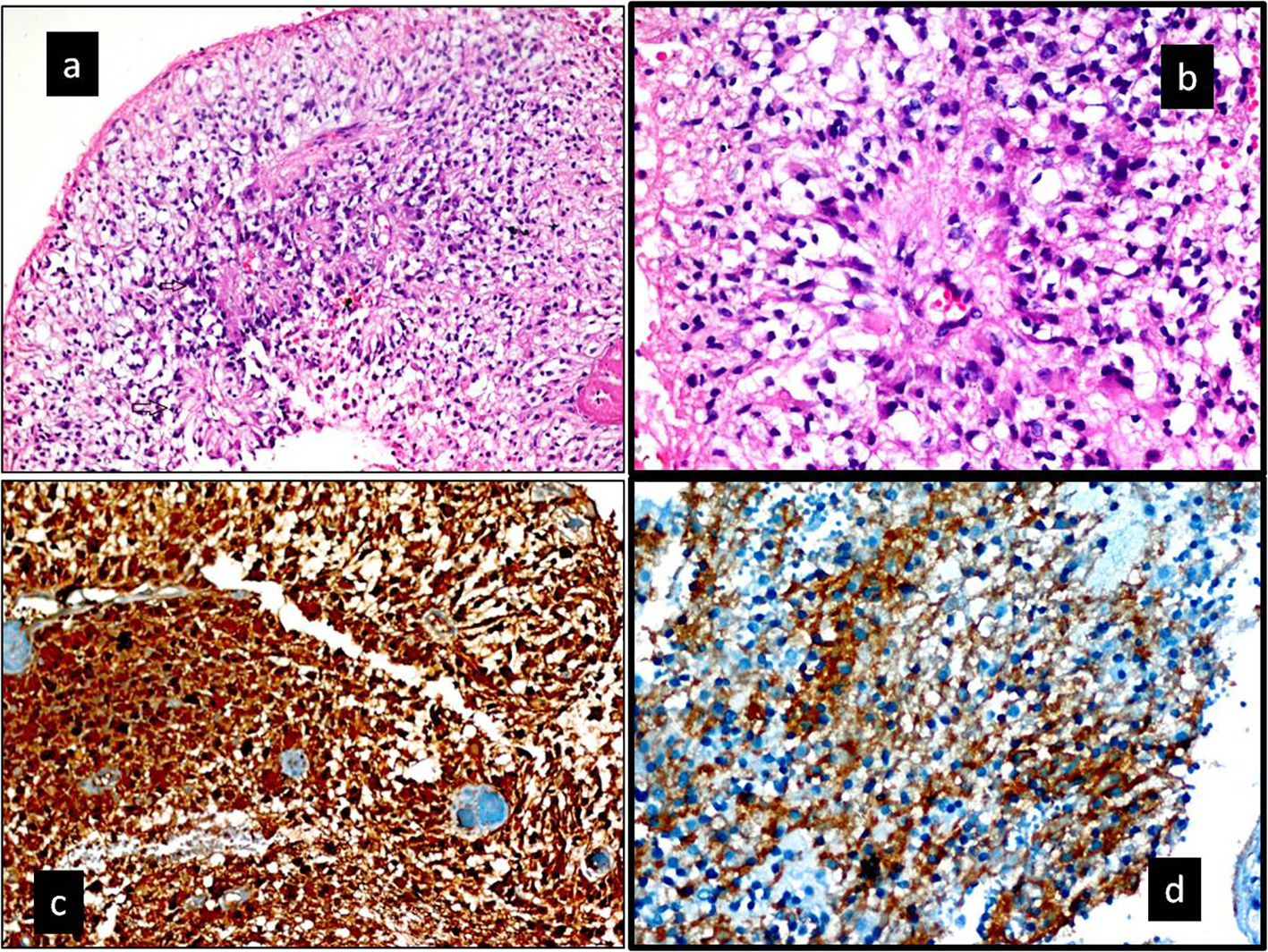
Figure 4. (a, b) Hematoxylin and eosin-stained section. (a) Increased cellularity with perivascualr pseudorosettes and a high mitotic index (black arrow) (× 200). (b) Pseudorosettes lined by pleomorphic cells with nuclear atypia and mitotic figures. Typical features for anaplastic ependymoma (H&E, × 400). (c, d) Immunohistochemical staining. (c) Tumor was diffuse positive with S-100 protein (× 200). (d) Tumor was diffuse positive with CD99 protein (× 400). Ki-67 labeling index in the anaplastic component was 75% in the tumor. In addition, tumor was diffuse positive with GFAP.

Tables

Table 1. Summary of 47 Cases With Supratentorial Cortical Anaplastic Ependymoma (WHO Grade 3) Before June 2014 in the Literature (No Connection to the Ventricles) [6, 8, 9, 12, 14-29]
 
Author, year and referenceAgeSexLocationPresentationNeuroimaging appearanceDifferent radiologic or clinics findings
M: male; F: female; C+: contrast enhancement; CAL: calcification; Het: heterogeneous; Hom: homogeneous.
1Vinchon, 2001 [25]15MTemporalHeadacheSolid + cystic
0.3MParietalSeizureSolid
13FTemporalSeizureSolid
11MParietalHeadacheSolid + cystic
2Takeshima, 2002 [26]70FFrontalLoss of consciousnessSolidRepeated intra-tumoral hemorrhage
3Kojima, 2003 [27]56FTemporalVertigo, apathieSolidMild CAL + hemorrhage
4Moritani, 2003 [22]50FTemporalHeadacheSolid
5Miyazawa, 2007 [28]33MParietalLoss of consciousnessSolidIntra-tumoral hemorrhage
6Niazi, 2009 [15]36FTemporalGeneralize seizureSolid, small cysts, Het C+
18MFrontoparietalFocal seizureSolid, Hom C+
7Alexiou, 2010 [18]10FFrontalHeadacheSolid, cystic, Het C+
8Hamano, 2010 [20]15MParietalHeadacheSolidMassive CAL
9Park, 2010 [23]17FFrontal (parafalcine)Headache, focal seizureSolidExtra-axial, mimicking meningioma
10Von Gompel, 2011 [21]12MParietalSeizuresMulti cystic
25MFrontalSeizuresMulti cystic
59MFrontalSeizuresSolidNon-cystic
11Davis, 2011 [6]22FFronto-temporalHeadache, dysarthriaSolid + cystic
12Kutlay, 2011 [9]11FFrontoparietalHeadacheSolid + cystic
13Ng, 2012 [16]51FBifrontalIncidentalSolid + small multicysticDiffuse CAL, presenting as a butterfly lesion
14Ohla, 2012 [24]29MParietalSeizureSolid
15Romero, 2012 [12]23MFrontalSeizureSolid, central necrotic, Het C+
16Singh, 2012 [29]35MFrontal (parafalcine)SeizureSolidMimicking meningioma
17Elsharkawy, 2013 [8]25MParietalHemiparesis, seizureSolidMimicking metastases
18Iwamoto, 2013 [17]61MTemporalHeadacheSolidIntra-tumoral hemorrhage
19Liu, 2013 [14] (21 cases)33 +/-1:01Frontal 11; parietal 5 frontoparietal 2; mix 3Seizure 45%Solid 60%
Solid + cystic
Usually Het C+
20Khilji, 2014 [19]10MFrontalHeadache focal seizureSolid
21Present case28FParietalHeadache, hemiparesis and focal seizureLarge pure cysticTogether pregnancy


Table 2. Immunohistochemical Findings of Brain Cortical Tumors [7, 17]
 
Tumor typeGFAPS-100 proteinVimentinCD99Pan-CK
The differential diagnosis was made according to the table with immunohistochemical findings. GFAP: glial fibrillary acidic protein; Pan-CK: pan-cytokeratin; R: rare.
Anaplastic ependymoma++-+-
Anaplastic meningioma-+(R)+--
Glioblastoma multiforme++---
Oligodendroglioma+(R)+---
Metastase of epithelial tumor----+


Table 3. Comparison of Pregnant Case With Anaplastic Ependymomas [44]
 
1Kaminski, 2003 [44]18FPosterior fossa in 4 ventricleStarted first trimesterHeadache, apathySolidOperated in first trimester
2Present case (2014)28FParietal cortexStarted third trimesterHeadache, hemiparesisPurely cysticOperated after birth