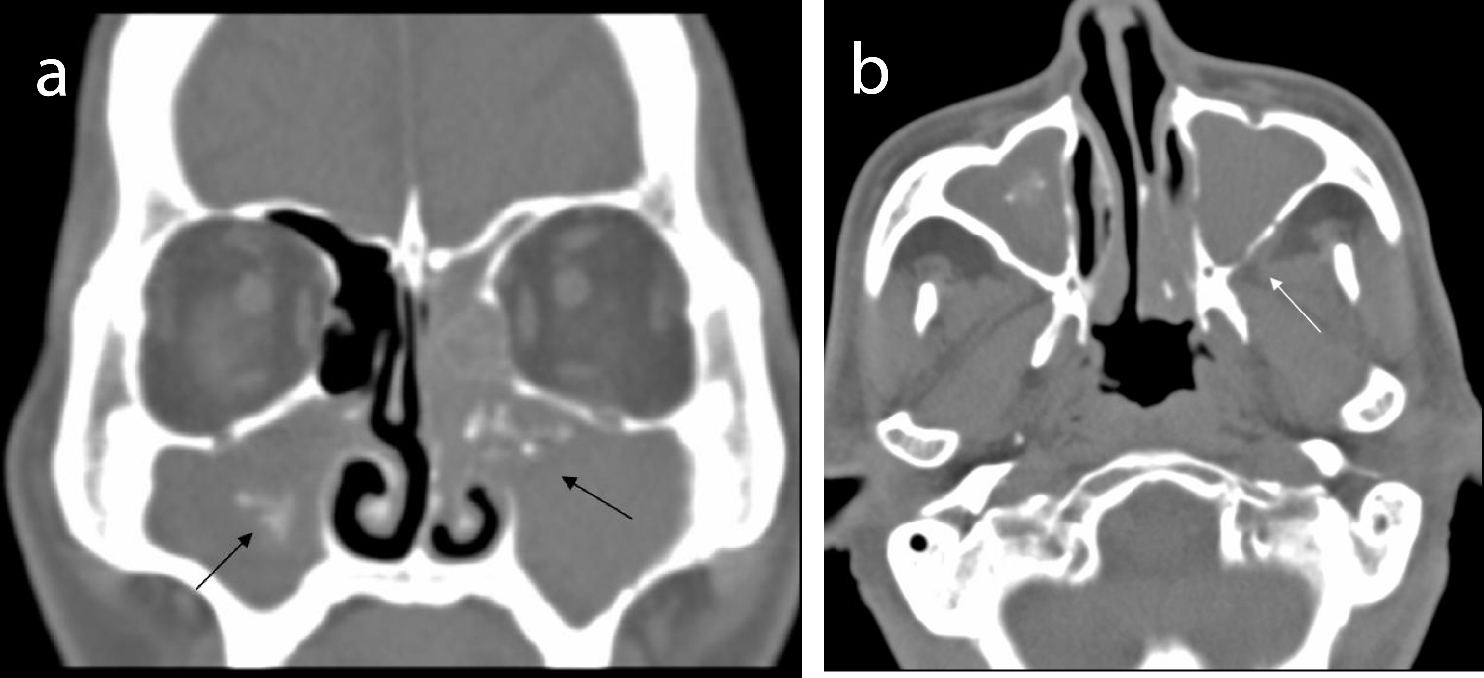
Figure 1. (a) Unrenced CT scan shows opacification of paranasal sinuses with intrasinus calcifications (black arrows) in bilateral maxillary sinuses. (b) Bony erosion (white arrow) over posterior wall of the left maxillary sinus.
| Journal of Medical Cases, ISSN 1923-4155 print, 1923-4163 online, Open Access |
| Article copyright, the authors; Journal compilation copyright, J Med Cases and Elmer Press Inc |
| Journal website http://www.journalmc.org |
Case Report
Volume 2, Number 4, August 2011, pages 162-165
Coexistence of Extramedullary Plasmacytoma and Fungus Balls in the Paranasal Sinuses: Report a Case
Figures