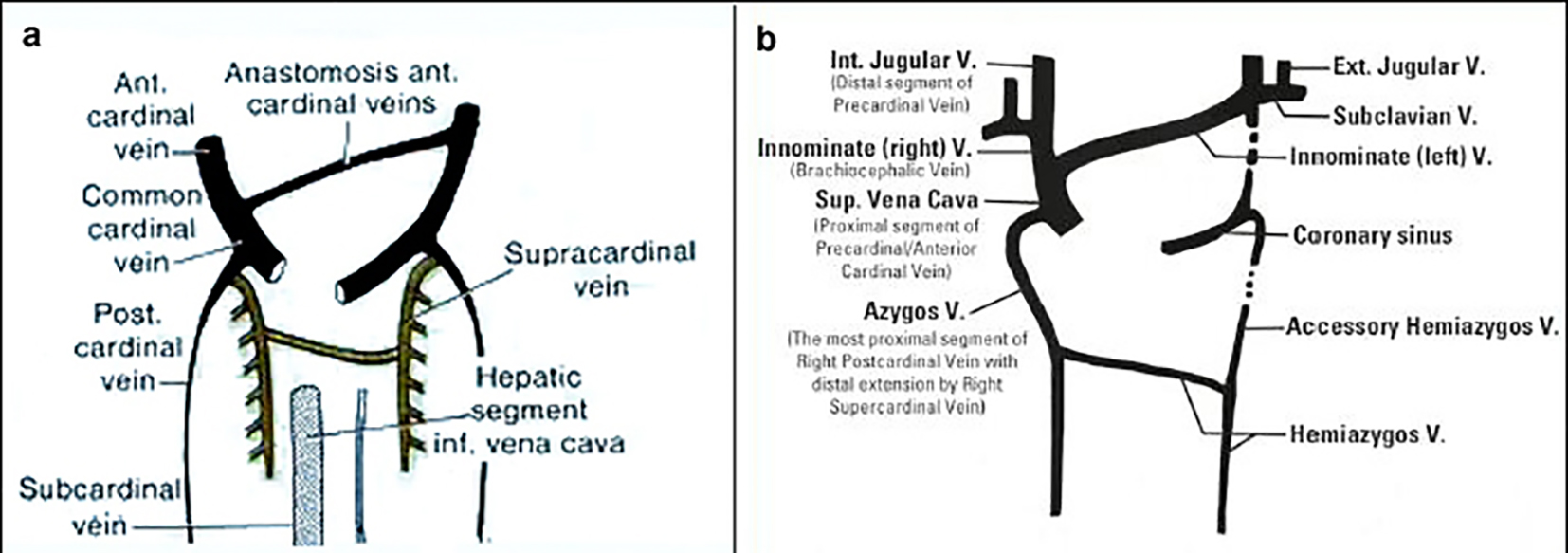
Figure 1. Precardinal/anterior cardinal vein developmental process (image taken from Phlebolymphology. Vol. 19, No. 4, 2012).
| Journal of Medical Cases, ISSN 1923-4155 print, 1923-4163 online, Open Access |
| Article copyright, the authors; Journal compilation copyright, J Med Cases and Elmer Press Inc |
| Journal website http://www.journalmc.org |
Case Report
Volume 7, Number 7, July 2016, pages 253-257
A Persistent Left-Sided Superior Vena Cava With Agenesis of the Right Superior Vena Cava in a Patient Who Underwent Liver Transplantation: A Case Report
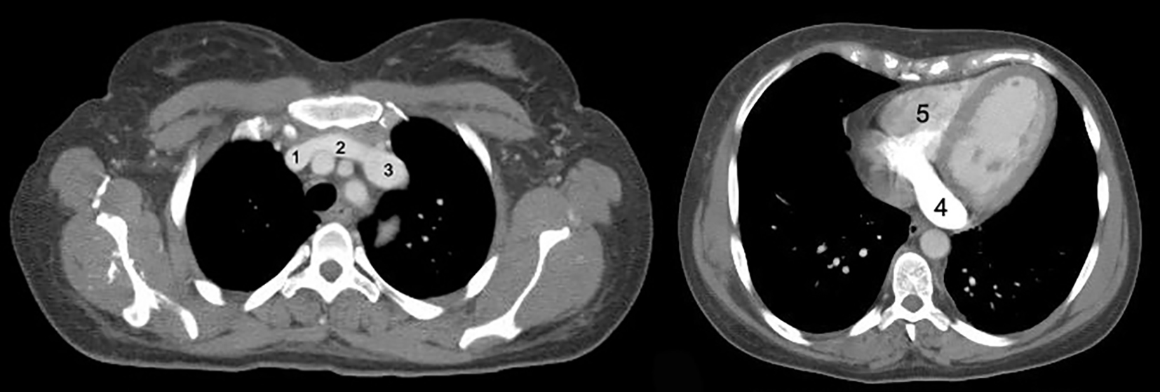
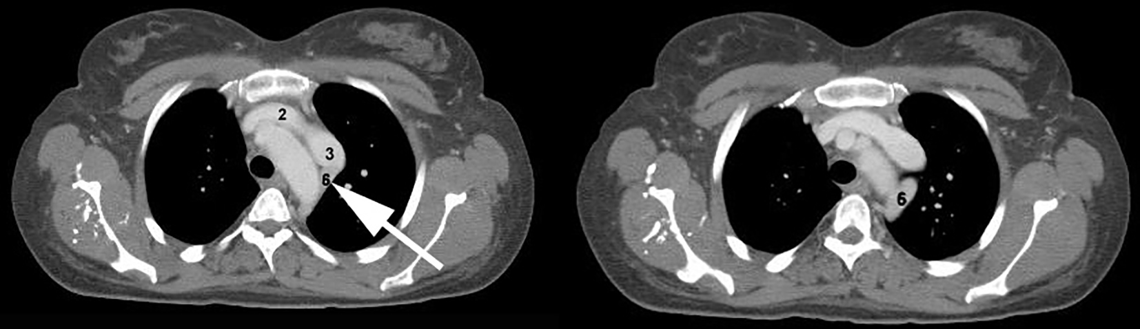
Figures