Journal of Medical Cases, ISSN 1923-4155 print, 1923-4163 online, Open Access
Article copyright, the authors; Journal compilation copyright, J Med Cases and Elmer Press Inc
Journal website http://www.journalmc.org

Case Report

Volume 2, Number 4, August 2011, pages 178-183


Report of a Case: Pseudoaneurysm of the Cystic Artery With Hemobilia Treated by Arterial Embolization

Figures

Figure 1.
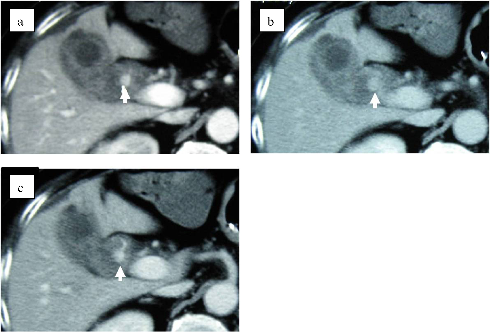
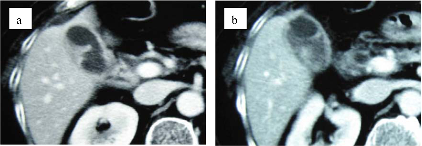
Figure 1. (a) Contrast-enhanced computed tomography shows thickening of gallbladder wall. (b) Contrast-enhanced CT shows enhanced lesion in the gallbladder. No structure of pseudoaneurysm is suspected.
Figure 2.
Figure 2. (a) MRI suspects two stones in the gall bladder (arrows). (b) MRCP suspects clots or stones in common bile duct (arrows).
Figure 3.
Figure 3. Endoscopy demonstrates blood oozing from the papilla of Vater (arrow).
Figure 4.
Figure 4. Dynamic CT shows 12 mm nodular lesion at the neck of gall bladder with an enhancement pattern parallel to the artery (arrows). (a) Arterial phase. (b) Balance phase. (c) Portal venous phase.
Figure 5.
Figure 5. (a) Selective right hepatic angiography shows a pseudoaneurysm (arrow) of the cyctic artery. (b) After embolization with microcoils, arterial inflow to the cystic artery is completely blocked.