Journal of Medical Cases, ISSN 1923-4155 print, 1923-4163 online, Open Access
Article copyright, the authors; Journal compilation copyright, J Med Cases and Elmer Press Inc
Journal website http://www.journalmc.org

Case Report

Volume 2, Number 6, December 2011, pages 292-295


Tumor Embolism Presenting as Rapidly-forming Cavitary Lesion

Figures

Figure 1.
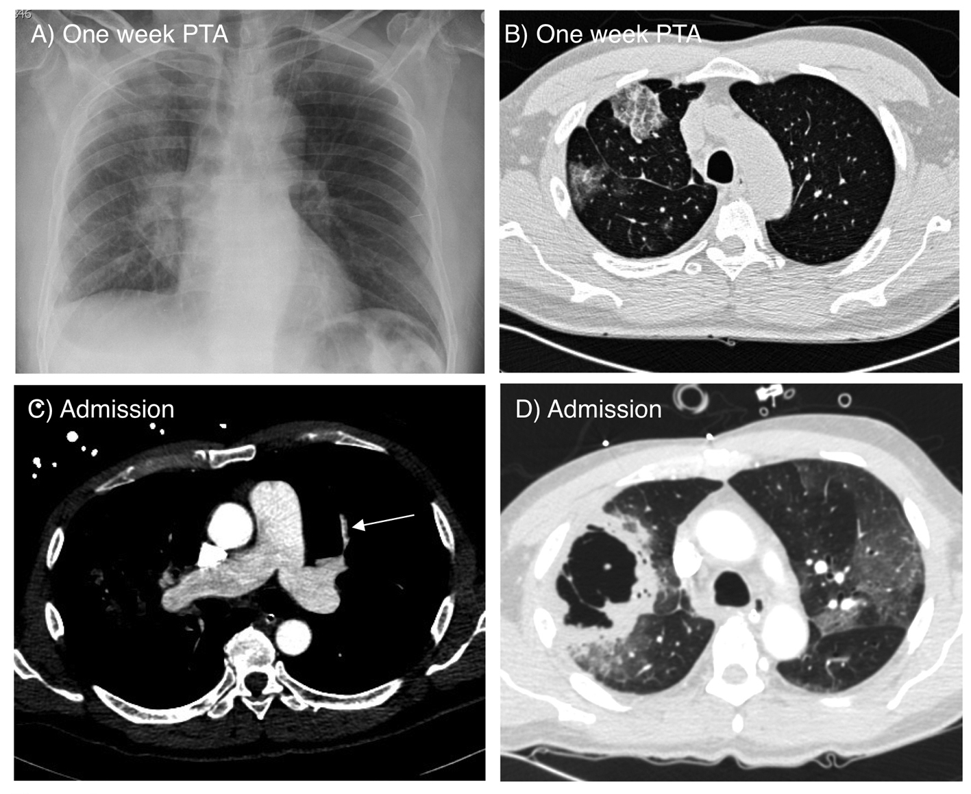
Figure 1. A) Chest radiograph on admission showing ground glass opacity and infiltrate in the right superior lung field and right perihilar area. B) Axial CT scan of the chest obtained one week prior to admission showing ground glass opacifications in the right middle and lower lobes. C) CT angiogram on admission showing a small embolus in a pulmonary artery branch of the left upper lobe (arrow). D) Lung window image of the CT angiogram demonstrating two adjacent large thick wall cavities in the right middle and lower lobes, corresponding to the ground glass opacities seen one week prior. There are also bilateral ground glass opacities.
Figure 2.
Figure 2. CT of the head showing hypodense lesions in the right frontal (long arrow) and cerebellar lobes (short arrow) consistent with embolic infarctions.
Figure 3.
Figure 3. Lung histopathology showing extensive tumor emboli. A) Low power image of right middle lobe with tumor embolus showing congested, but viable tissue to the left of the embolus and necrotic cavitary abscess to the right (*) (40 X). B) Higher power image of arterial and lymphatic/venous tumor emboli adhering to the endothelium and obstructing the vascular luminae (200 X).