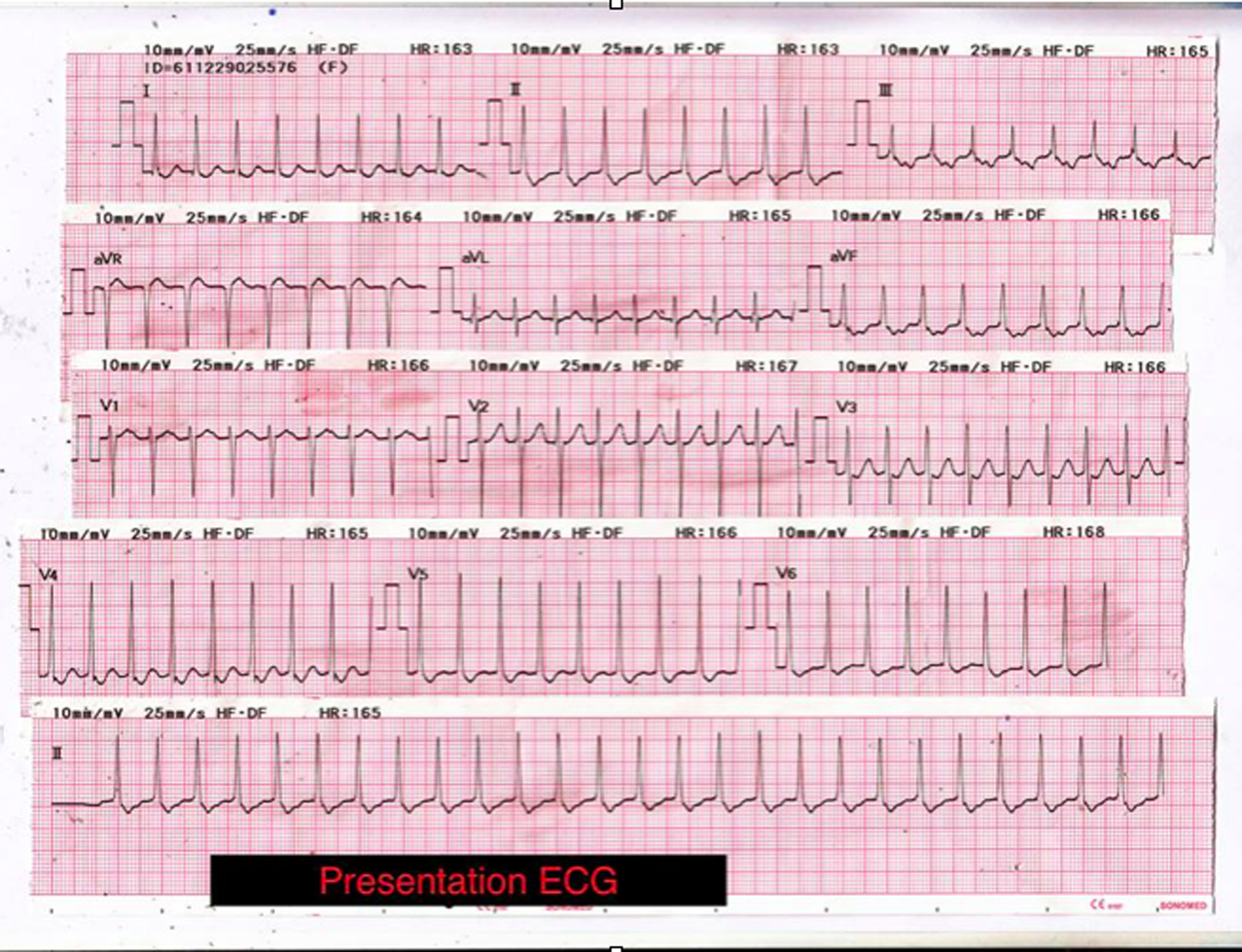
Figure 1. ECG upon arrival shows regular narrow complex tachycardia beating at a rate of 160 beats/min. Axis is normal with no acute ST segment changes. Non-specific ST segment depression and T wave inversion can be seen over the infero-lateral leads. Inverted P waves may be visible de-forming the ST segment indicating that atrial depolarisation occurs later than ventricular depolarisa-tion. In a patient with a narrow complex tachycardia the presence of such late P waves is frequently the only ECG evidence that the patient has an accessory pathway rather than a much more common atrioventricular nodal re-entrant tachycardia (AVNRT).