Journal of Medical Cases, ISSN 1923-4155 print, 1923-4163 online, Open Access
Article copyright, the authors; Journal compilation copyright, J Med Cases and Elmer Press Inc
Journal website http://www.journalmc.org

Case Report

Volume 7, Number 12, December 2016, pages 550-553


A Case of Peripartum Cardiomyopathy Affected by Left Ventricle Thrombus

Figures

Figure 1.
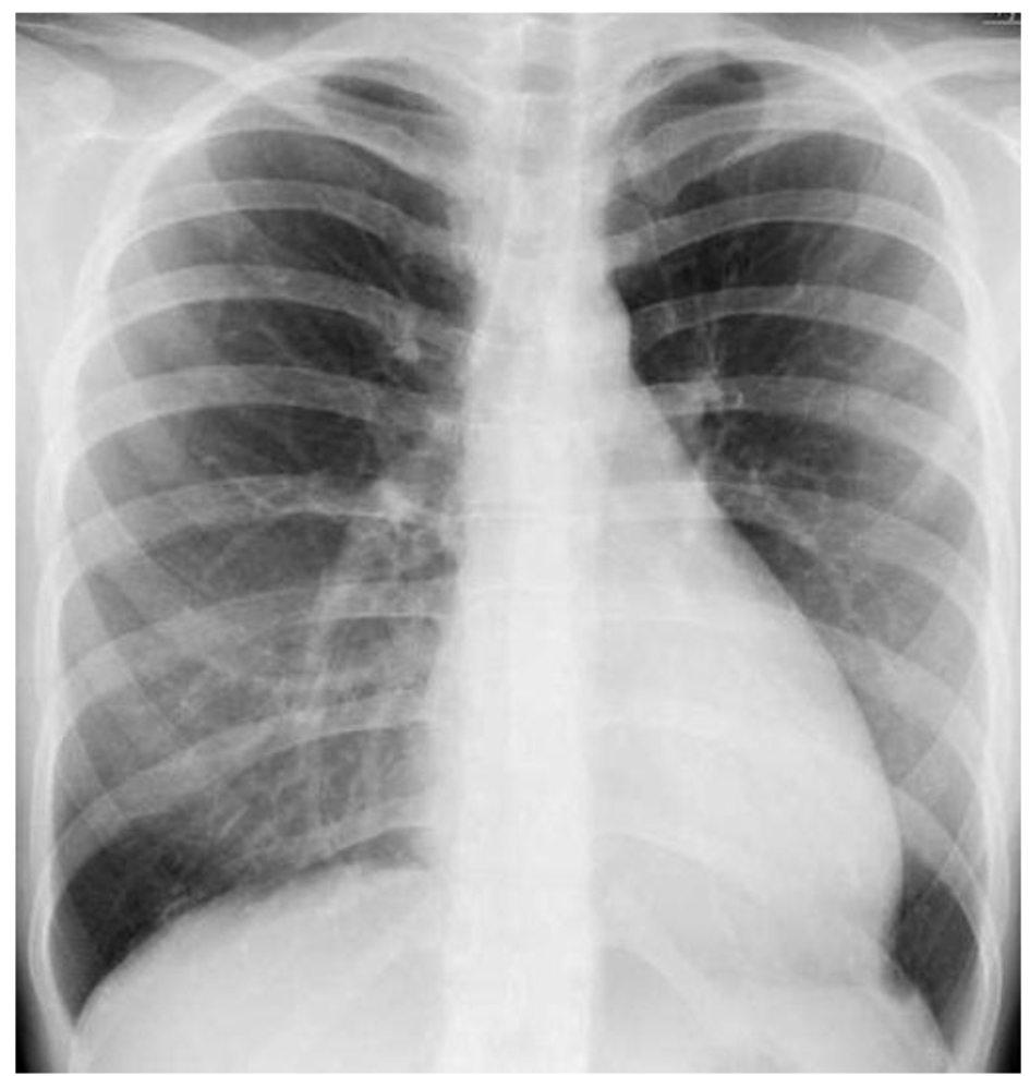
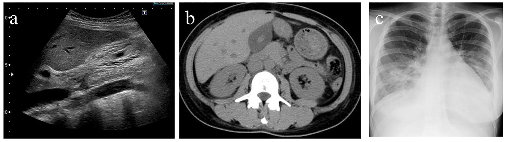
Figure 1. (a) Ultrasonography revealed an edematous gallbladder. (b) Non-contrast computed tomography revealed an edematous gallbladder, dilated hepatic and inferior jugular veins. (c) Chest X-ray revealed cardiomegaly with a cardio-thoracic ratio of 58%, pulmonary congestion, and pleural effusion.
Figure 2.
Figure 2. Transthoracic echocardiogram revealed a dilated left ventricle of 63 mm in diastolic phase (a) and 58 mm in systolic phase (b) with a reduced EF of 17%.
Figure 3.
Figure 3. Chest X-ray revealed disappearance of heart congestion and cardiomegaly on day 6.
Figure 4.
Figure 4. Transthoracic echocardiogram revealed thromboses of 16 × 8 (a) mm and 29 × 11 mm (b) in the patient’s left ventricle on day 10.