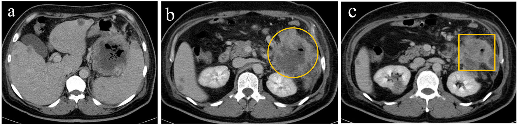
Figure 1. Contrast-enhanced computed tomography revealed a marked thickening of the gastric walls along with multiple liver metastases (a), a cystic mass surrounded by contrast-enhanced margins (circle) (b), and thickening of the walls in the transverse colon (square) (c).
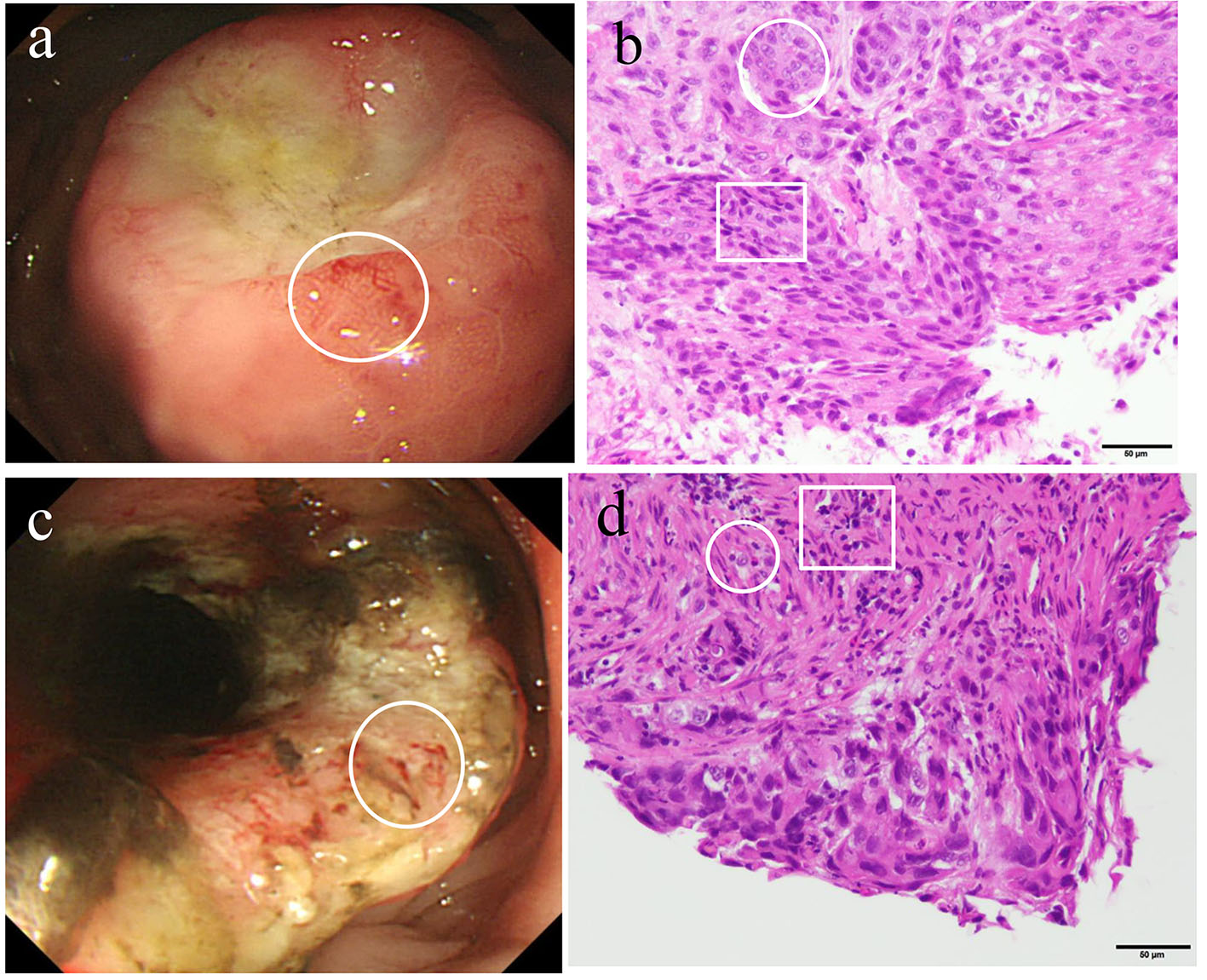
Figure 3. Upper gastric endoscopy revealed a raised mass with central depressive lesions in the middle body of the greater curvature (a). A biopsied specimen of the gastric tumor (circle) revealed the presence of both adenocarcinoma (circle) and squamous cell carcinoma (square) (b). A colonoscopy revealed a half-circumferential lesion in the splenic flexure of the transverse colon (c). A biopsied specimen (circle) revealed the presence of both adenocarcinoma (circle) and squamous cell carcinoma (square) (d).
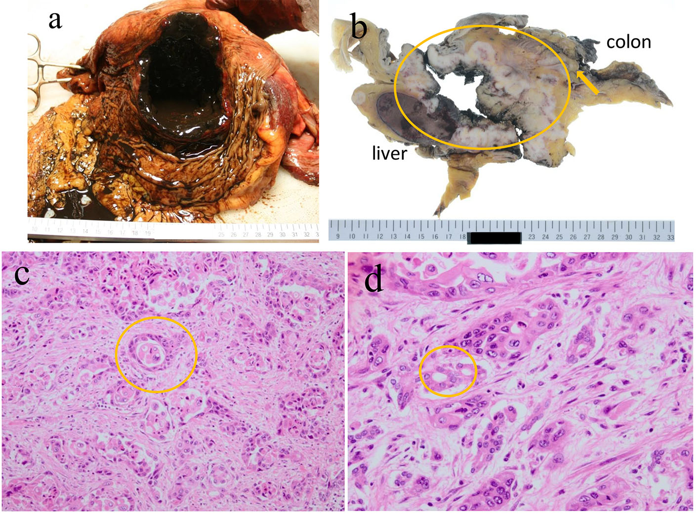
Figure 4. An autopsy revealed that the stomach was perforated and contained 100 mL of blood (a). The perforation site between the pancreatic tumor in the tail and the transverse colon (arrow indicates perforation site; circle indicates the pancreatic tumor) (b). Histological findings of pancreatic tumors (c) and invaded gastric lesions (d) revealed atypical cells accompanied by inflammatory cells and prolific fibrosis, making an alveolar and sheet-like structure, along with keratinization, an intercellular bridge, and a glandular formation.