Figures
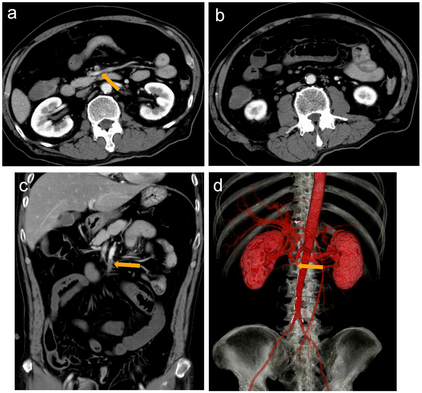
Figure 1. Contrast-enhanced computed tomography (CT) reveals a suspected thrombus (arrow) in the main trunk of the superior mesenteric artery (a) and dilated small intestines (b). Coronal section (c) and three-dimensional contrast-enhanced CT (d) suggest that the thrombus is located in the main trunk of SMA at the level of the second branch of the jejunal artery (arrow).
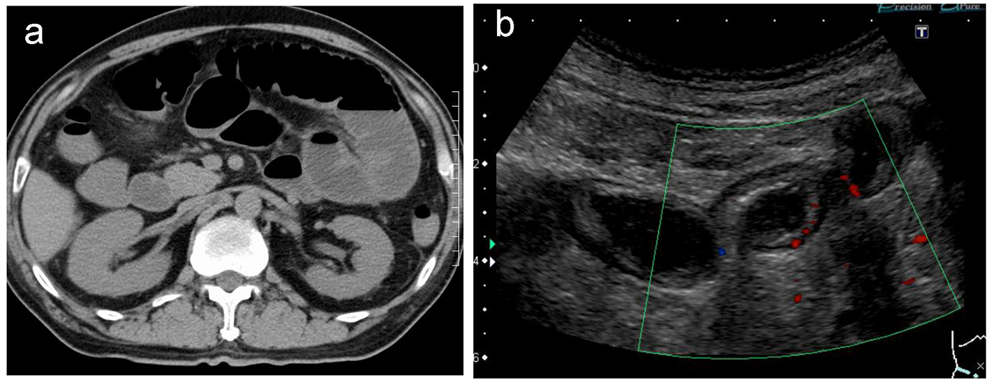
Figure 2. Non-contrast computed tomography reveals extensively dilated small intestines (a). Doppler ultrasonography reveals edematous and dilated small intestines with reduced intestinal perfusion (b).
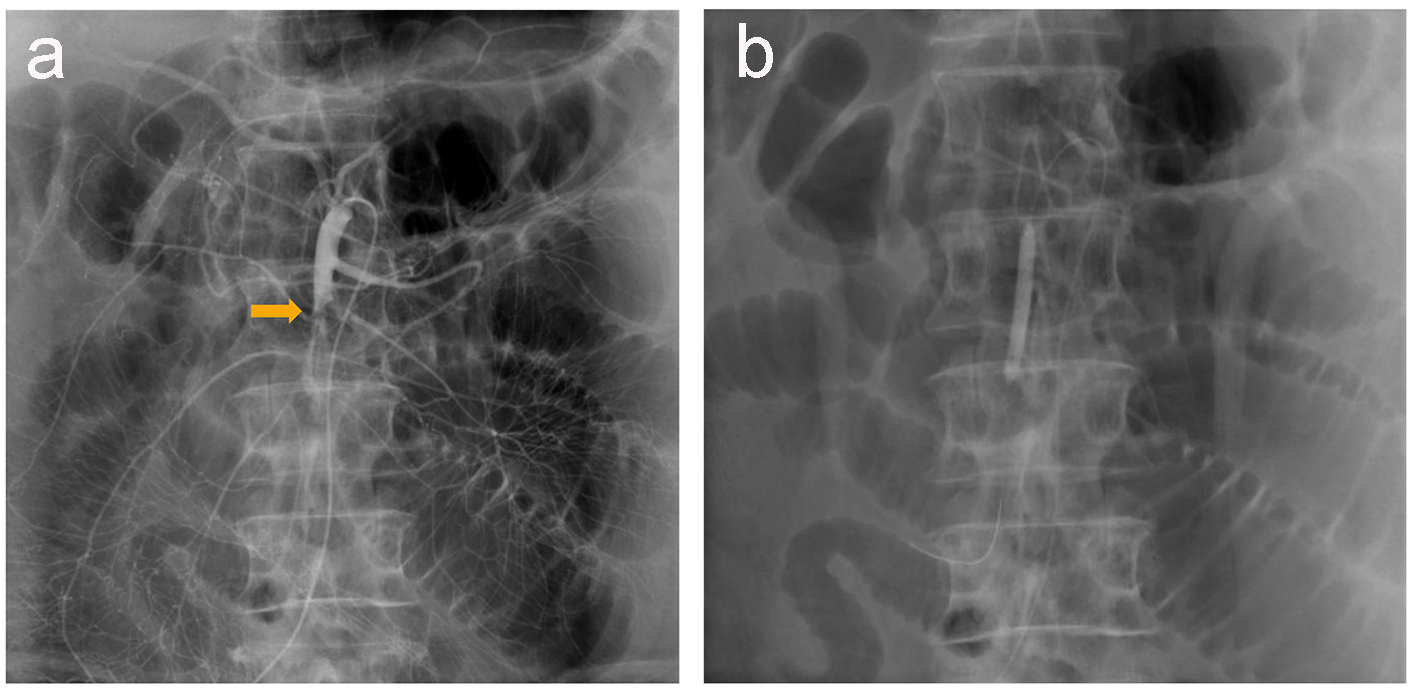
Figure 3. Abdominal angiography reveals a thrombus in the main trunk of SMA at the level of the second branch of the jejunal artery, with preserved peripheral circulation because of developed collateral arteries (a). A 0.019-inch guide wire and a balloon catheter are successfully delivered to the lesion (b).
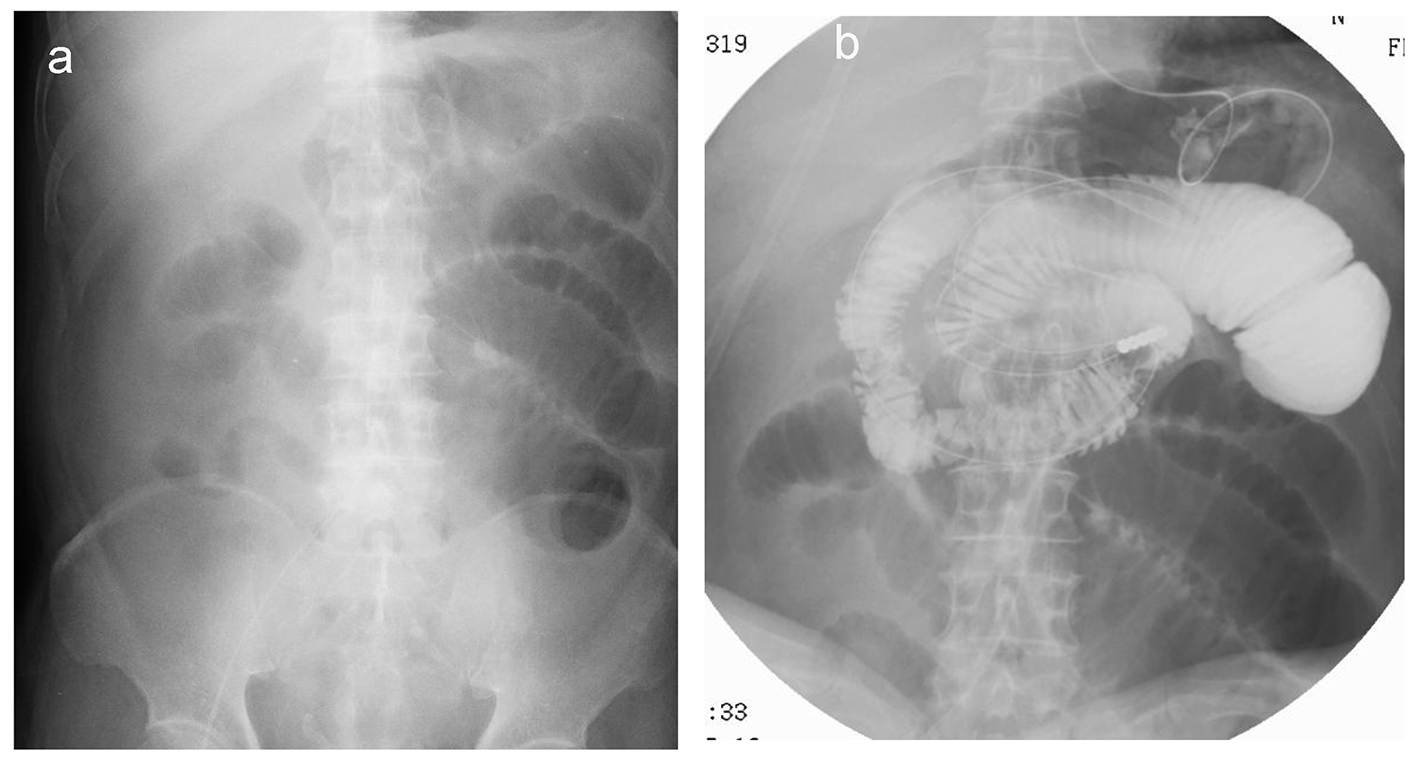
Figure 4. X-ray reveals dilated small bowels (a) that require placement of an ileus tube to deflate the bowel pressure (b).
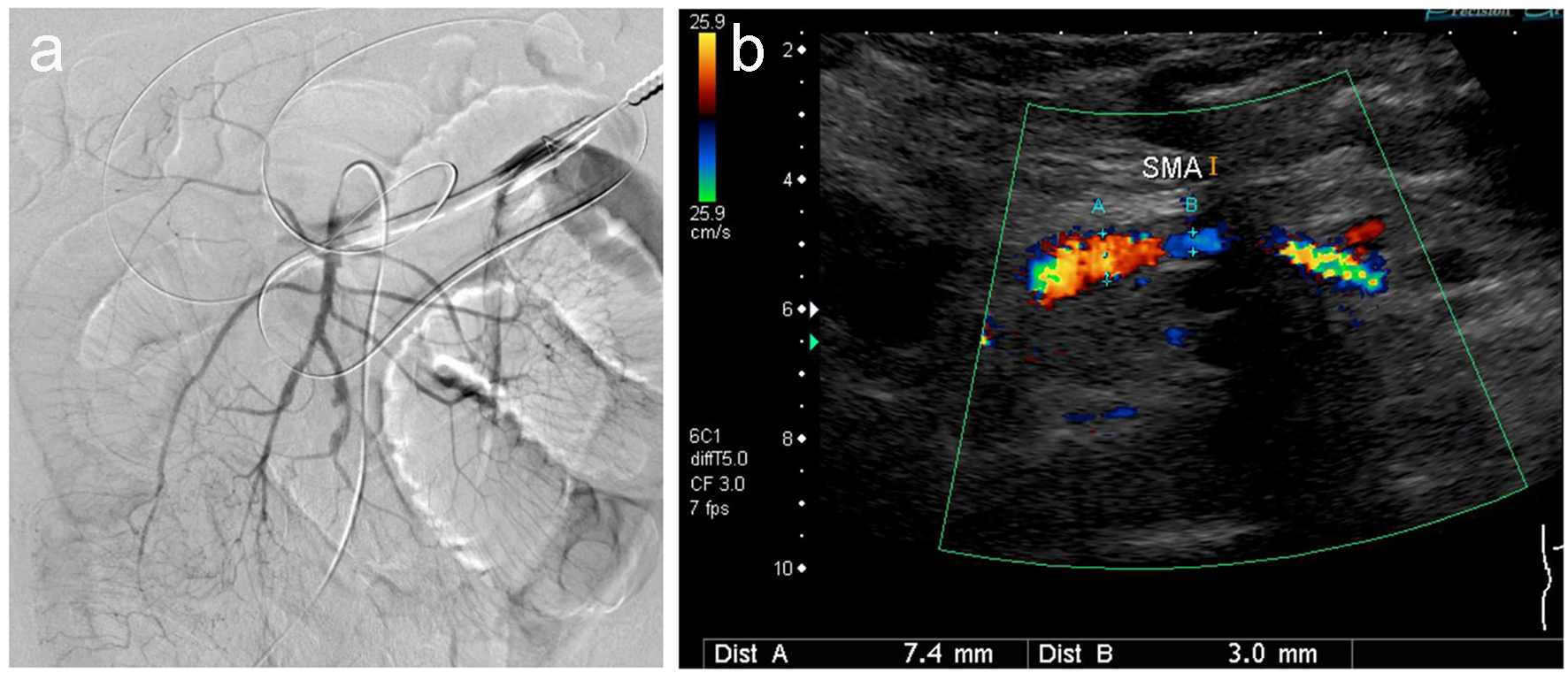
Figure 5. Angiography (a) and ultrasonography (b) revealed no thrombus, but irregular stenotic arteries are evident.