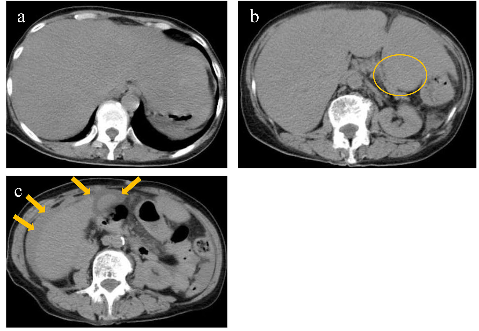
Figure 1. Non-contrast computed tomography revealed diffusely swollen liver with obscure vessel structures (a), a mass in the pancreatic tail (circle) (b), and ascites around the gall bladder and liver (arrows) (c).
| Journal of Medical Cases, ISSN 1923-4155 print, 1923-4163 online, Open Access |
| Article copyright, the authors; Journal compilation copyright, J Med Cases and Elmer Press Inc |
| Journal website http://www.journalmc.org |
Case Report
Volume 8, Number 5, May 2017, pages 141-144
Ruptured Anaplastic Pancreatic Carcinoma With Liver Metastasis: A Case Report
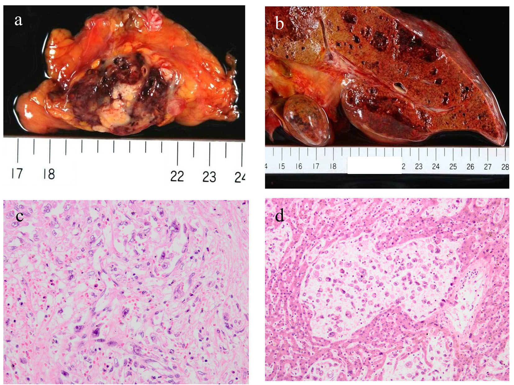
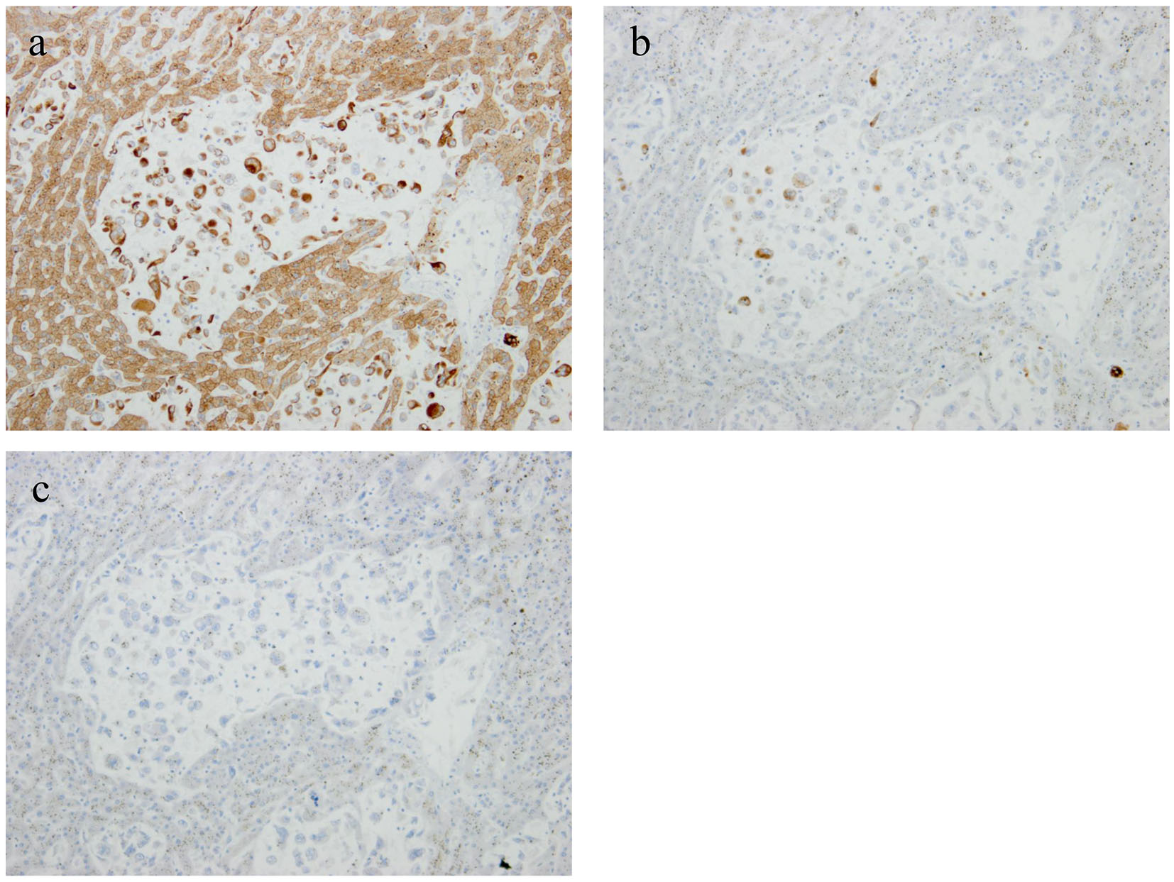
Figures