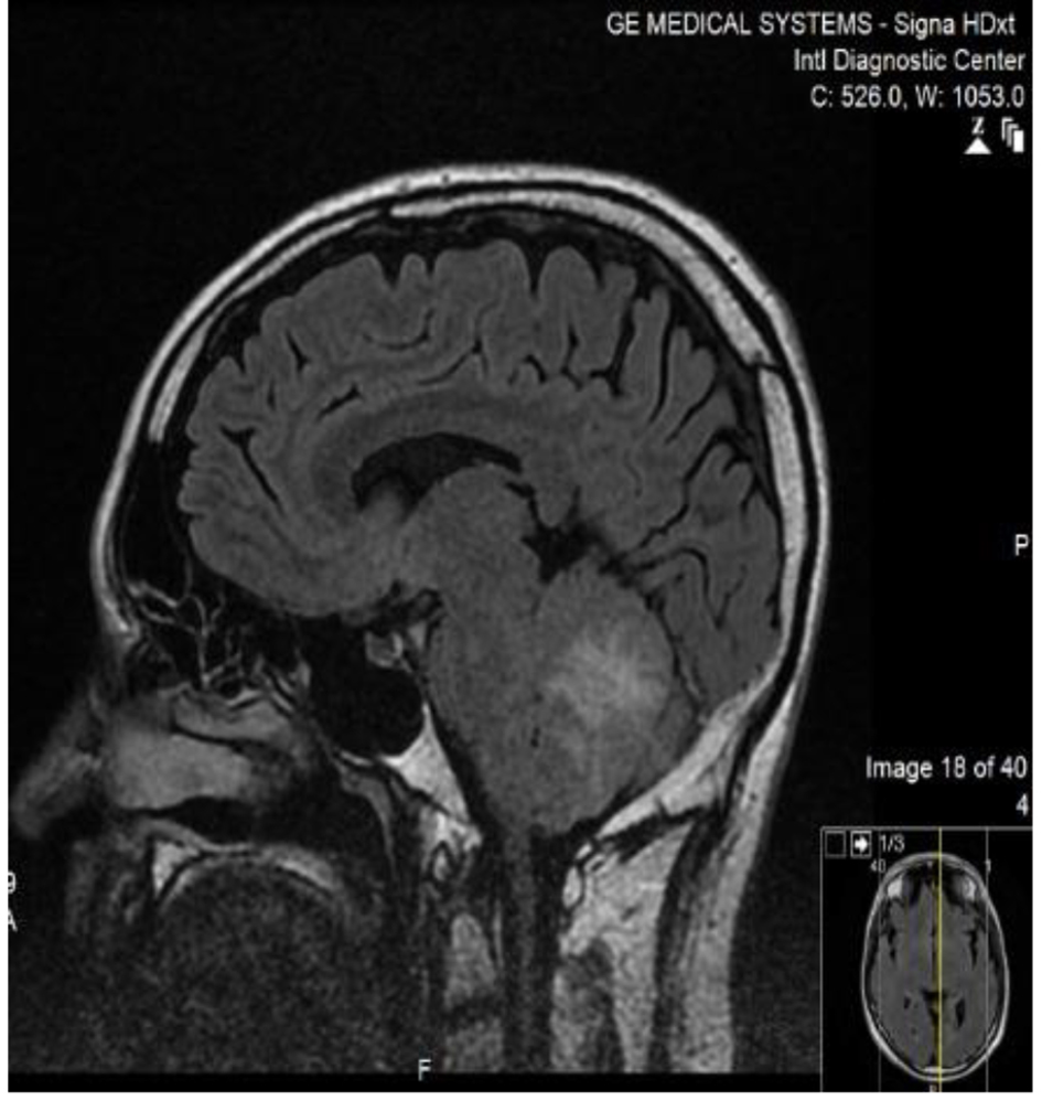
Figure 1. MRI brain/FLAIR sequence of the patient sagittal cut, showing high signal intense lesion in the middle cerebellum, with surrounding edema.
| Journal of Medical Cases, ISSN 1923-4155 print, 1923-4163 online, Open Access |
| Article copyright, the authors; Journal compilation copyright, J Med Cases and Elmer Press Inc |
| Journal website http://www.journalmc.org |
Case Report
Volume 8, Number 8, August 2017, pages 249-251
Cerebellar Tumor Like Lesion in a Patient With Neurosarcoidosis
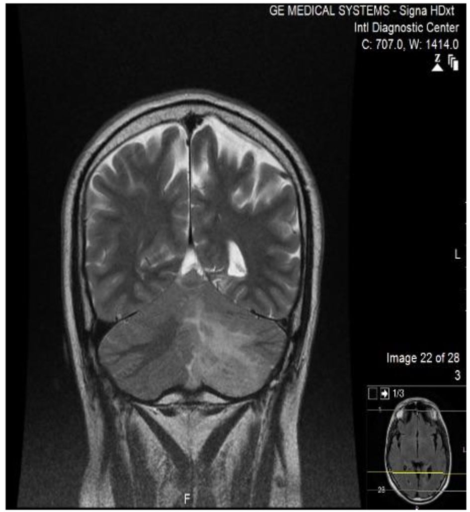
Figures
Table
| WBC | 2 cells |
| Protein | 0.54 g/L |
| Glucose | 6.6 mmol/L |
| PCR mycobacterium | Negative |
| Gram stain | Negative |
| Bacterial culture | Negative |
| Cytology | Lymphocytes |