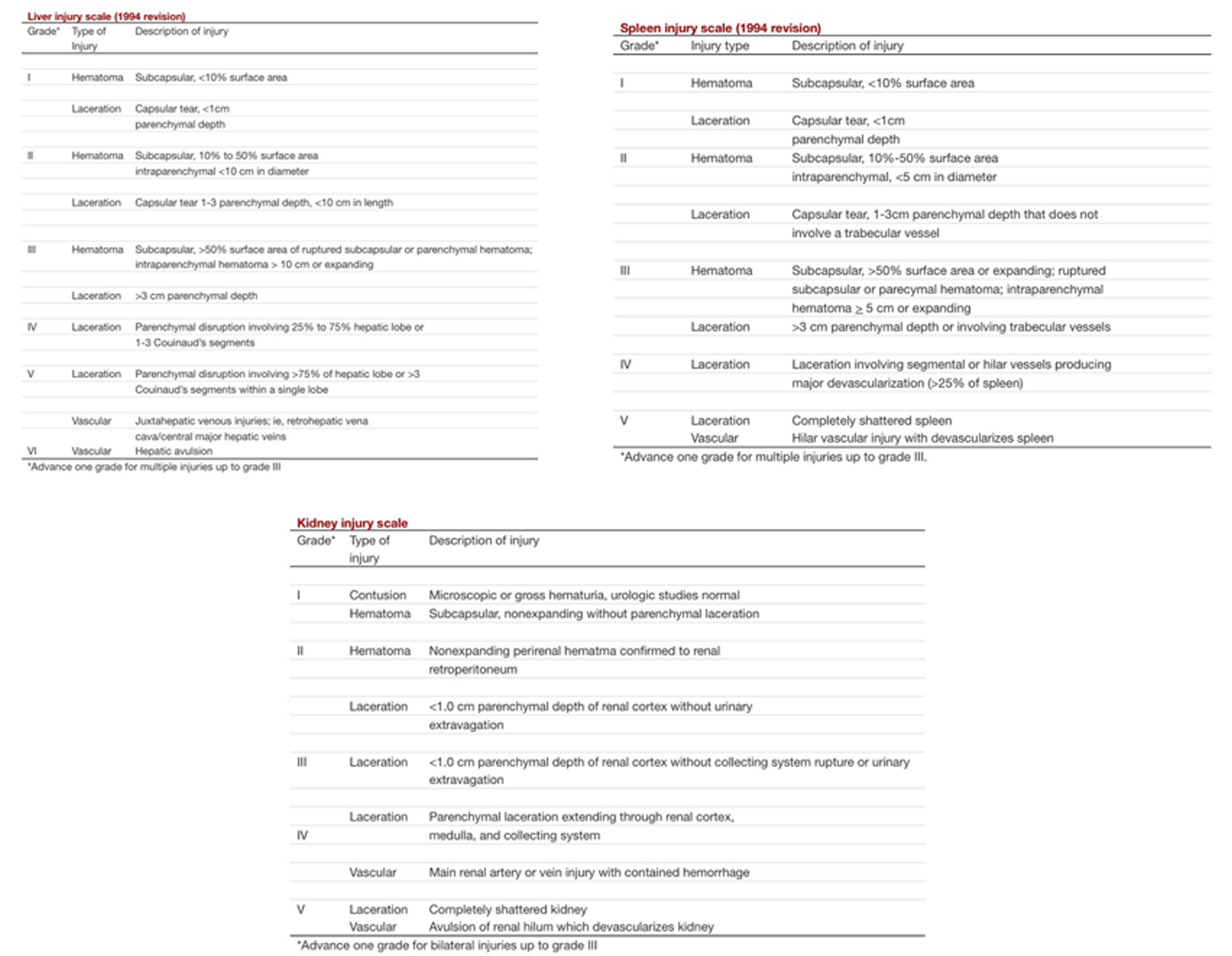
Figure 1. AAST grading of liver, spleen and renal injuries.
| Journal of Medical Cases, ISSN 1923-4155 print, 1923-4163 online, Open Access |
| Article copyright, the authors; Journal compilation copyright, J Med Cases and Elmer Press Inc |
| Journal website http://www.journalmc.org |
Case Report
Volume 8, Number 11, November 2017, pages 340-346
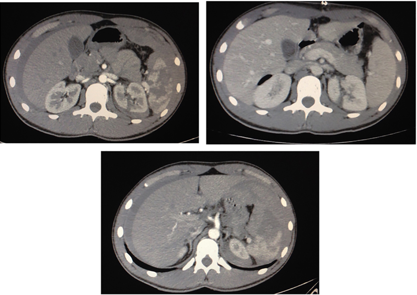
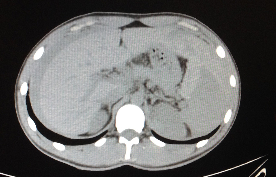
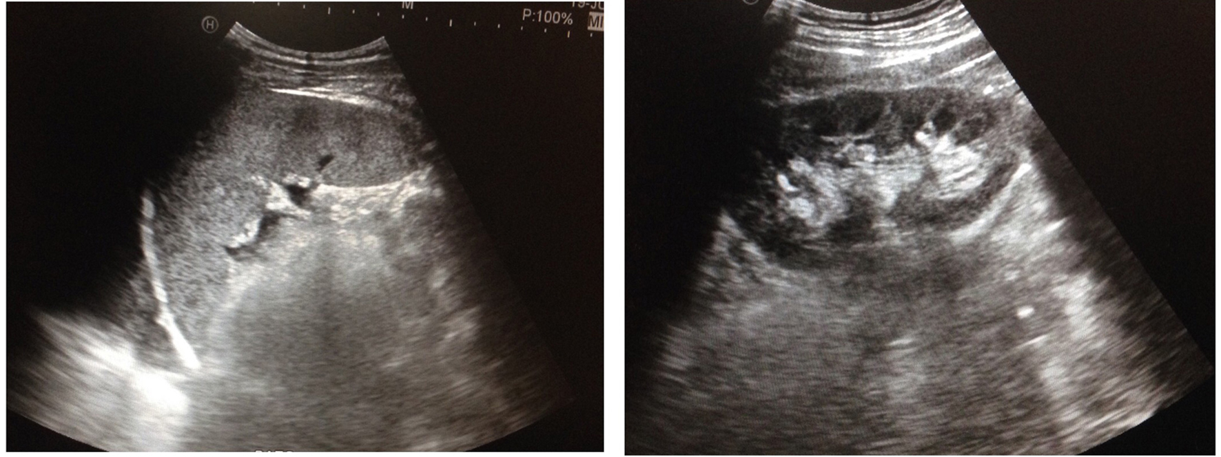
The Challenge of Blunt Abdominal Trauma in Children: Report of a Case and Review of Management
Figures