Journal of Medical Cases, ISSN 1923-4155 print, 1923-4163 online, Open Access
Article copyright, the authors; Journal compilation copyright, J Med Cases and Elmer Press Inc
Journal website http://www.journalmc.org

Case Report

Volume 9, Number 2, February 2018, pages 45-51


Intramuscular Cysts at the Proximal Calf: Two Case Reports and a Systematic Review of Literature

Figures

Figure 1.
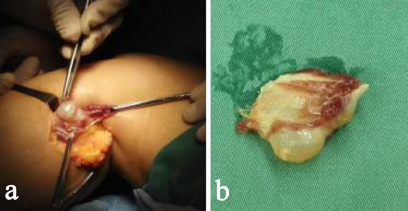
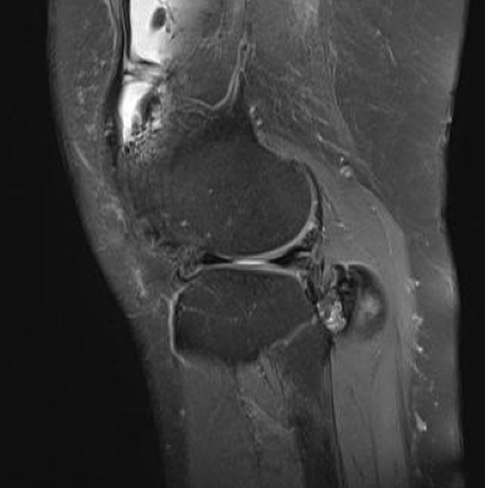
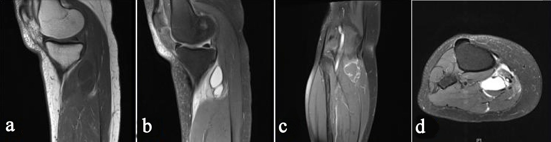
Figure 1. Preoperative magnetic resonance image of the right knee. Sagittal T1-weighted image (a), sagittal T2-weighted image (b), coronal T2-weighted image (c), and axial T2-weighted image (d) reveal a mass in the gastrocnemius medialis muscle, which does not communicate with the articular joint.
Figure 2.
Figure 2. Intra-operative cyst located in the gastrocnemius medialis muscle (a) and partial tissue of the excised cyst (b), as the cyst ruptured partly when excised.
Figure 3.
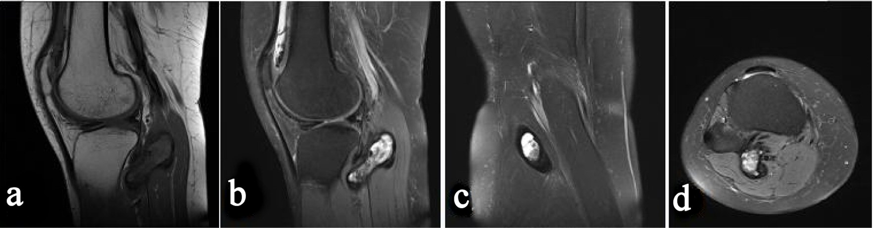
Figure 3. Preoperative magnetic resonance image (MRI) of the right knee. Sagittal T1-weighted image (a), sagittal T2-weighted image (b), coronal T2-weighted image (c), and axial T2-weighted image (d) reveal a mass in the gastrocnemius lateralis muscle. Thickened synovium with low signal intensity on both T1-weighted and T2-weighted images is found in the knee joint.
Figure 4.
Figure 4. Preoperative magnetic resonance image (MRI) of the right knee. Sagittal T2-weighted image showed the cyst wall to be attached to the knee joint capsule.