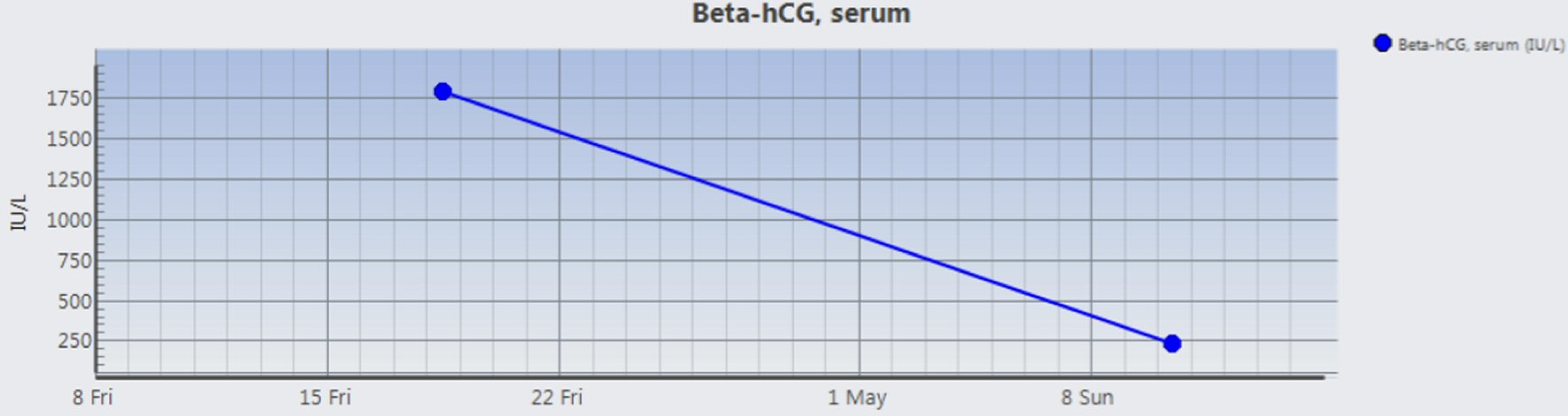
Figure 1. Postpartum trend of β-hCG levels.
| Journal of Medical Cases, ISSN 1923-4155 print, 1923-4163 online, Open Access |
| Article copyright, the authors; Journal compilation copyright, J Med Cases and Elmer Press Inc |
| Journal website http://www.journalmc.org |
Case Report
Volume 9, Number 1, January 2018, pages 34-36
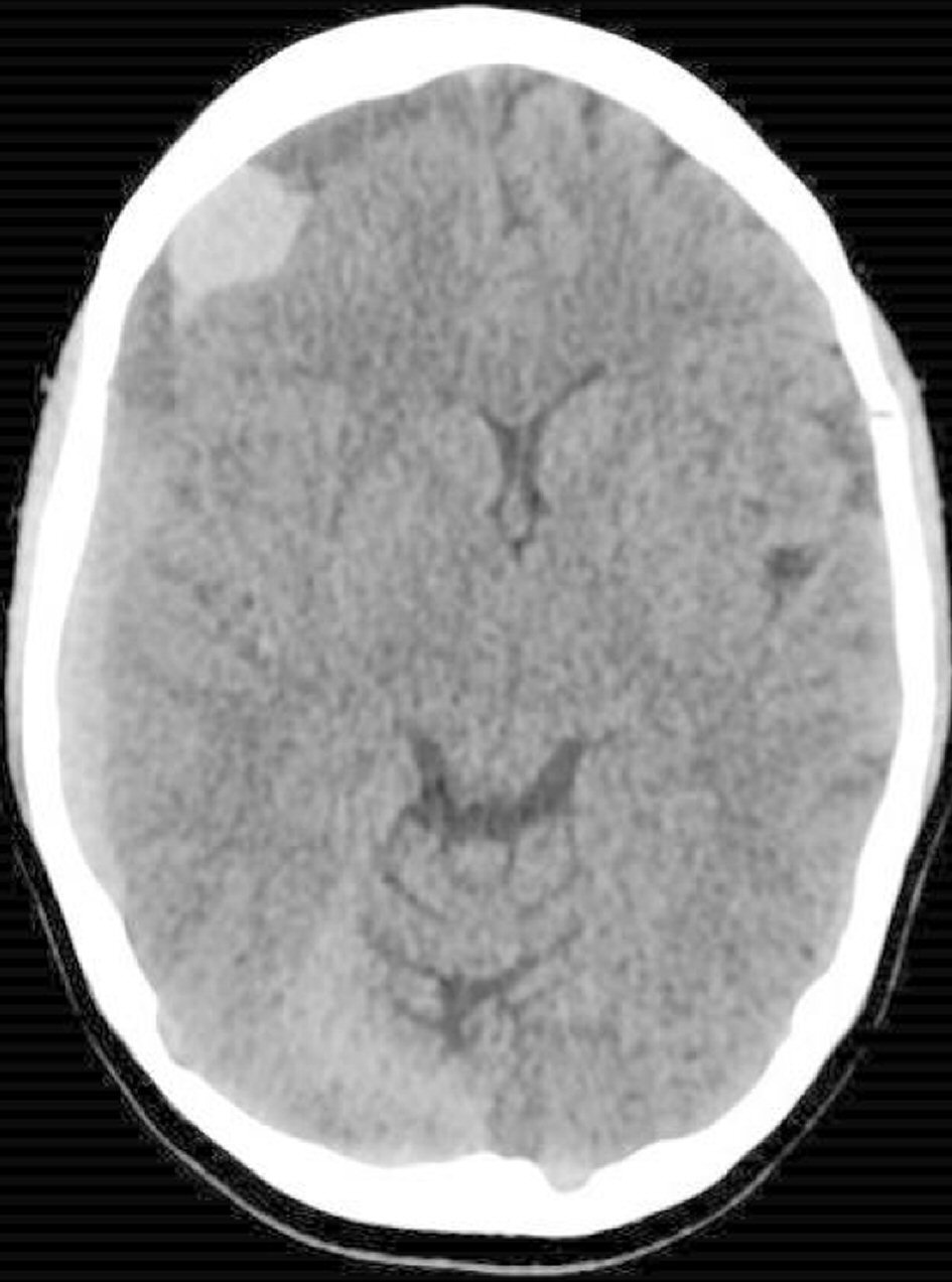
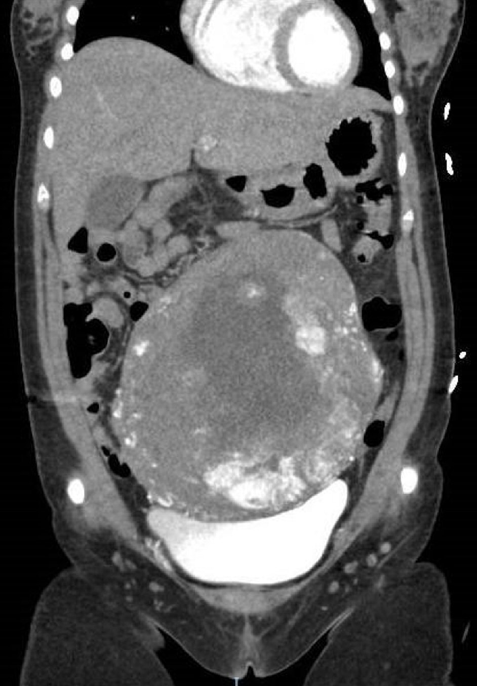
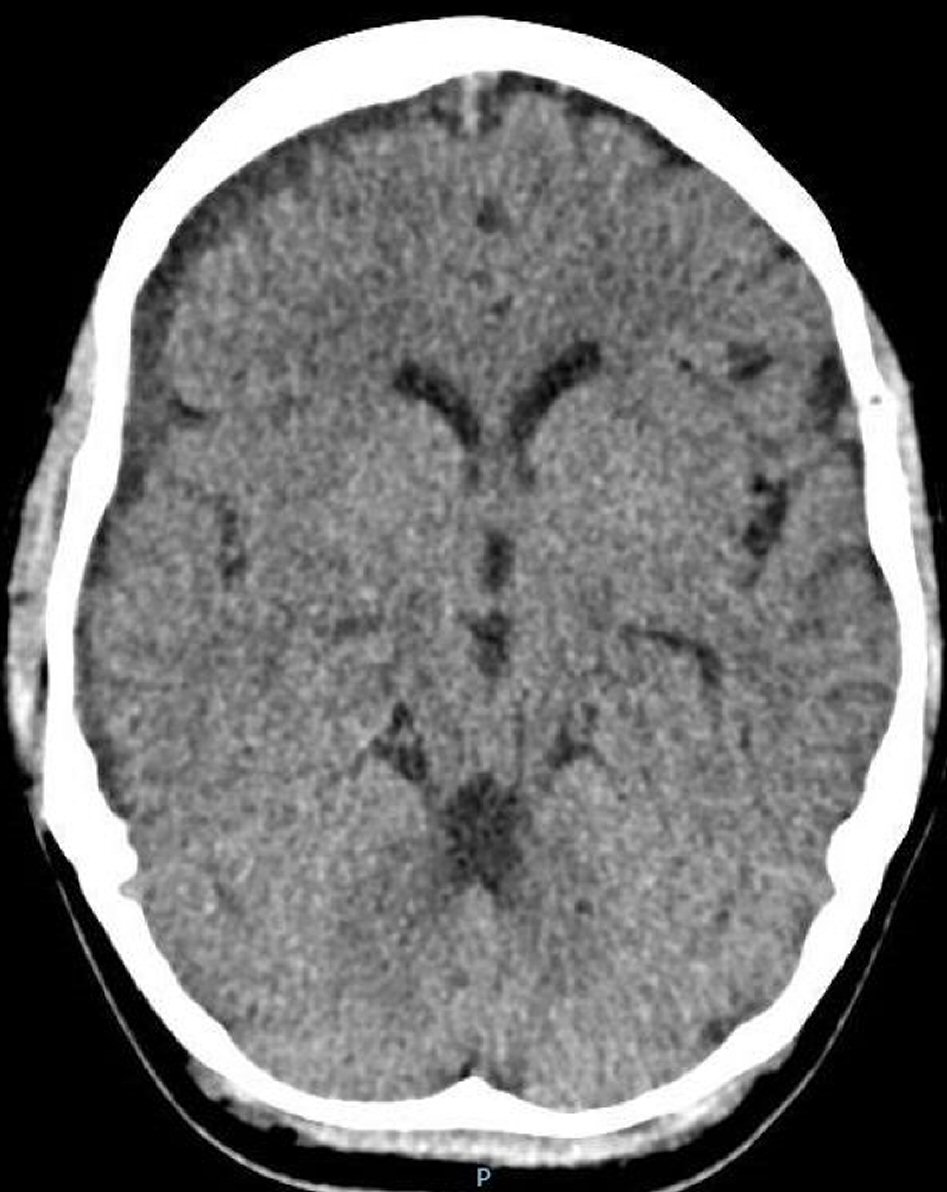
An Unusual Presentation of Acute Subdural Hematoma Secondary to Disseminated Intravascular Coagulation Following Conservative Management of Placenta Increta
Figures
Tables
| WBC: white blood cell; PTT: partial thromboplastin time; PT: prothrombin time. | |
| Hemoglobin | 11.5 g/dL |
| WBC count | 6.19 × 10⁹/L |
| Platelet | 146 × 109/L |
| PTT | 39.2 s |
| PT | 16.8 s |
| Fibrinogen | 0.49 g/L |
| D-dimer | > 32.00 mg/L FEU |
| WBC: white blood cell; PTT: partial thromboplastin time; PT: prothrombin time. | |
| Hemoglobin | 9.3 g/dL |
| WBC count | 8.05 × 10⁹/L |
| Platelet | 184 × 109/L |
| PTT | 26.2 s |
| PT | 10.1 s |
| Fibrinogen | 3.19 g/L |