Journal of Medical Cases, ISSN 1923-4155 print, 1923-4163 online, Open Access
Article copyright, the authors; Journal compilation copyright, J Med Cases and Elmer Press Inc
Journal website http://www.journalmc.org

Case Report

Volume 9, Number 3, March 2018, pages 86-89


Nuclear Protein of the Testis Midline Carcinoma

Figures

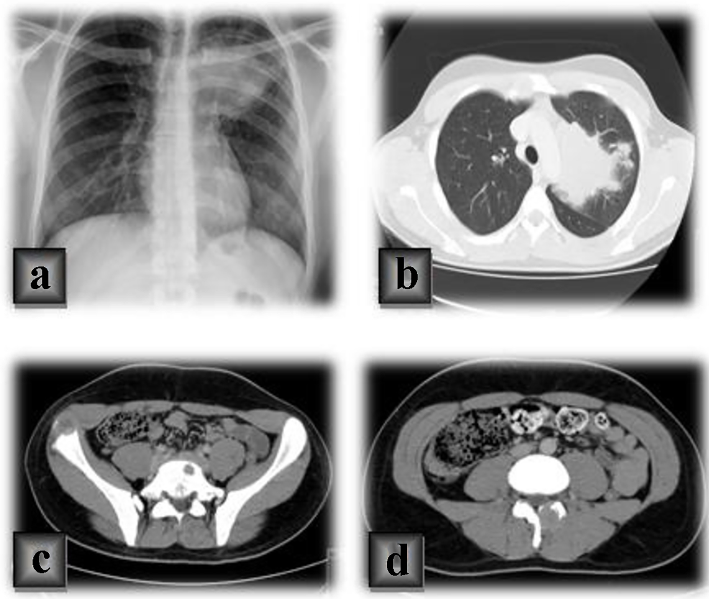
Figure 1.
Figure 1. (a) Chest radiograph of a large mediastinal hypotransparency on the left side. (b) A chest computed tomography (CT) of a pulmonary mass in the left upper pulmonary lobe, in contact with the aorta and the left pulmonary artery, mediastinal lymph nodes and bone metastases in the sternum and left costal arch. (c, d) CT scan of secondary lesions in the spine and iliac bone in the abdomen and pelvis.
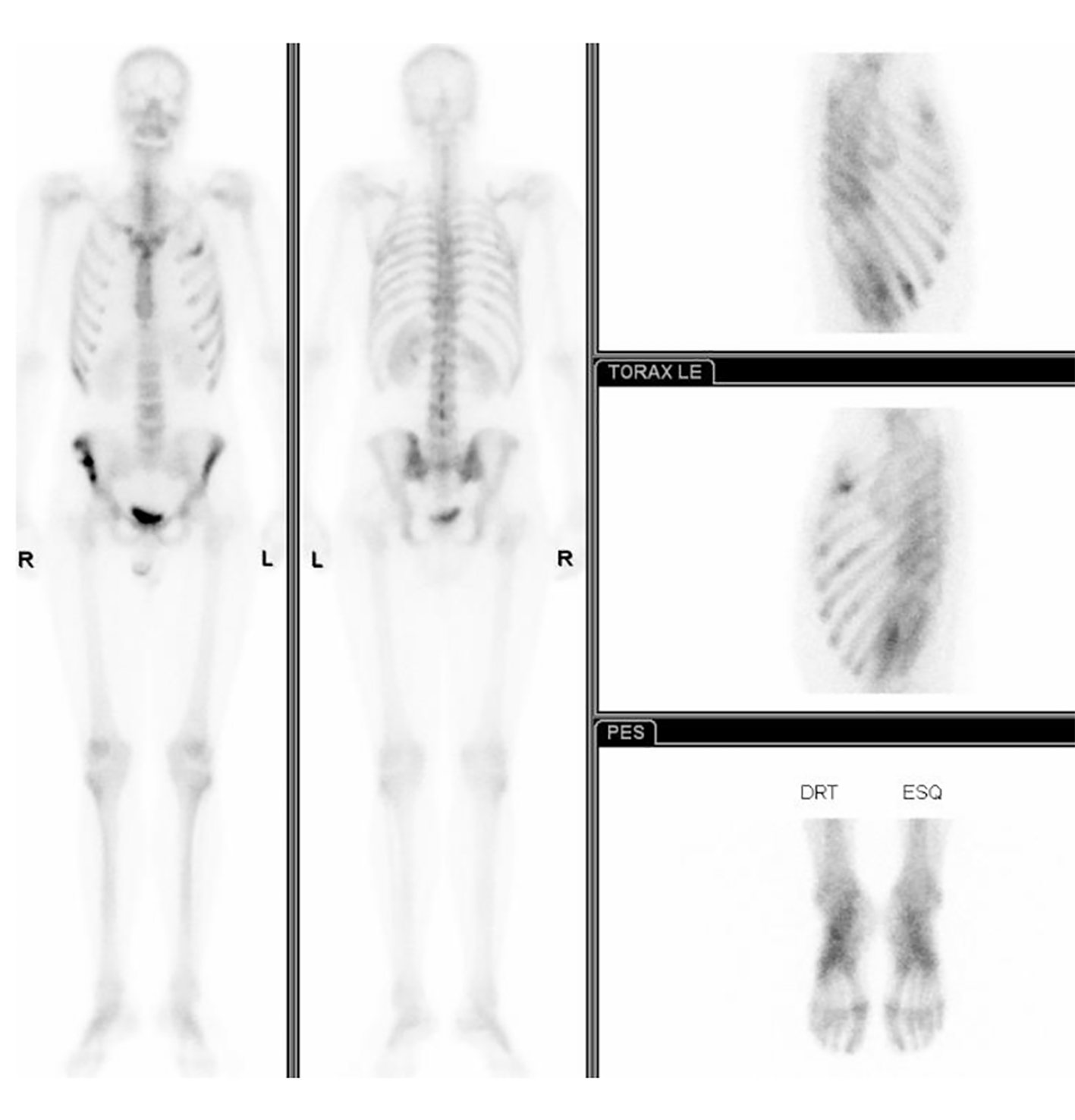
Figure 2.
Figure 2. The bone scintigraphy showing pathological foci in bilateral iliac bone, sternum, second and fifth left costal arches and the right lesser trochanter.