Journal of Medical Cases, ISSN 1923-4155 print, 1923-4163 online, Open Access
Article copyright, the authors; Journal compilation copyright, J Med Cases and Elmer Press Inc
Journal website http://www.journalmc.org

Case Report

Volume 9, Number 5, May 2018, pages 131-134


A Case of Cecal Cancer With Heterotopic Ossification

Figures

Figure 1.
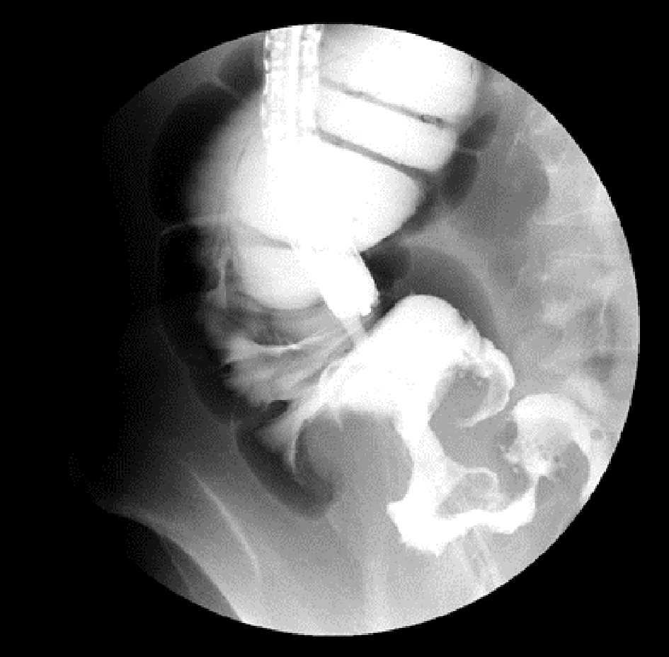
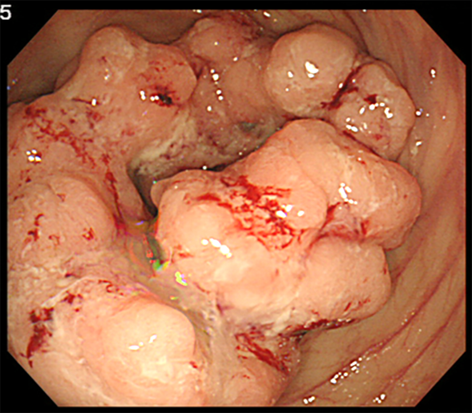
Figure 1. Colonoscopy revealing a cecal cancer with bleeding.
Figure 2.
Figure 2. Irrigoscopy showing the typical apple-core sign at the cecum.
Figure 3.
Figure 3. Computed tomography scan showing an irregular cecal wall thickening without any sign of calcification or ossification.
Figure 4.
Figure 4. The resected specimen.
Figure 5.
Figure 5. (a) A section of the main tumor comprising moderately differentiated adenocarcinoma tissue. (b) A section of the white solid lesion showing heterotopic bone formation without dysplasia.