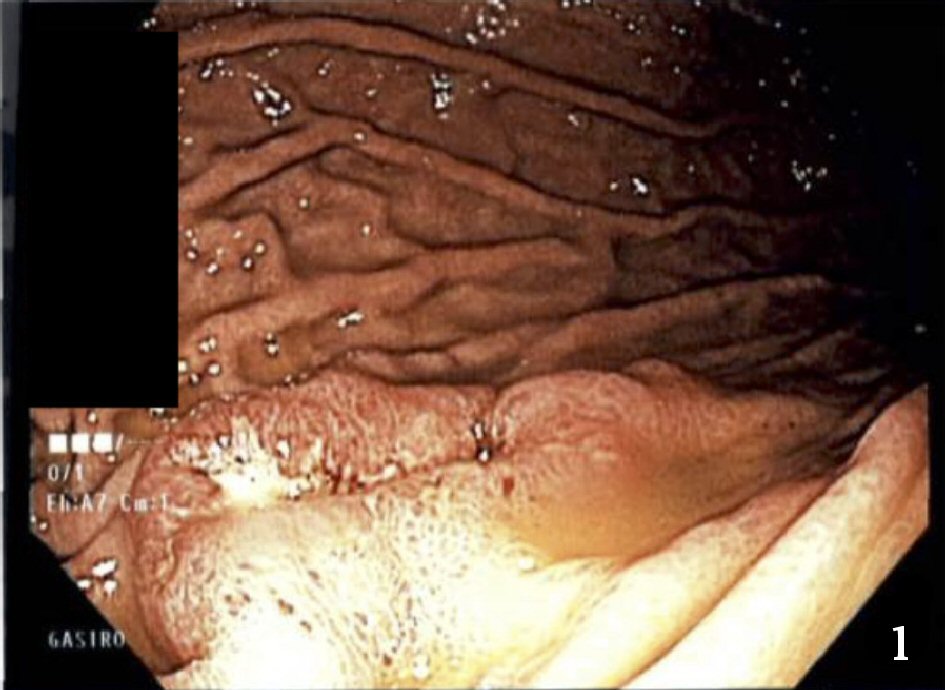
Figure 1. Photograph of stomach ulcer taken during upper endoscopy.
| Journal of Medical Cases, ISSN 1923-4155 print, 1923-4163 online, Open Access |
| Article copyright, the authors; Journal compilation copyright, J Med Cases and Elmer Press Inc |
| Journal website http://www.journalmc.org |
Case Report
Volume 9, Number 6, June 2018, pages 160-163
A Rare Presentation of Large Cell Neuroendocrine Carcinoma of the Lung
Figures