Journal of Medical Cases, ISSN 1923-4155 print, 1923-4163 online, Open Access
Article copyright, the authors; Journal compilation copyright, J Med Cases and Elmer Press Inc
Journal website http://www.journalmc.org

Case Report

Volume 9, Number 6, June 2018, pages 173-176


Wellens’ Syndrome: An Atypical Presentation of an Already Silent Killer

Figures

Figure 1.
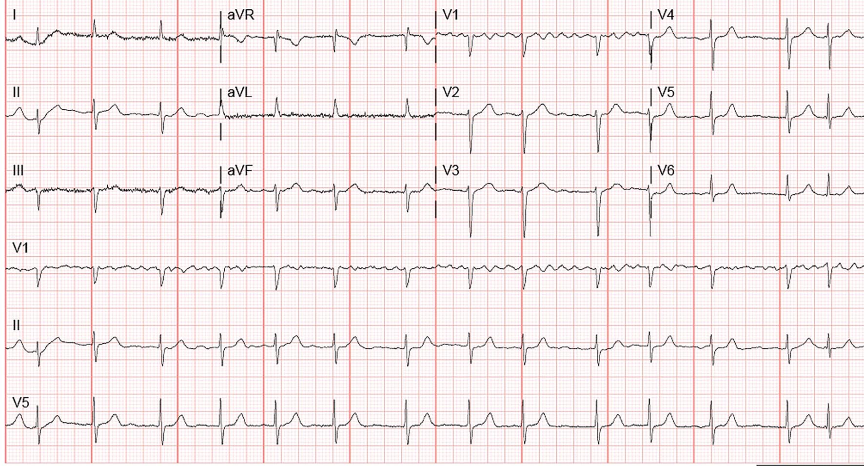
Figure 1. Admission EKG showing atrial fibrillation.
Figure 2.
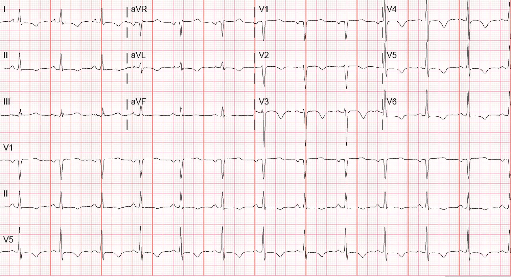
Figure 2. EKG performed after dynamic changes noted on telemetry showing sinus rhythm with rate of 75 beats per minute with left axis deviation, T-wave inversions in lead V2 to V6, I and aVL and less than 1-mm ST-segment depression in V6 and lead I.
Figure 3.
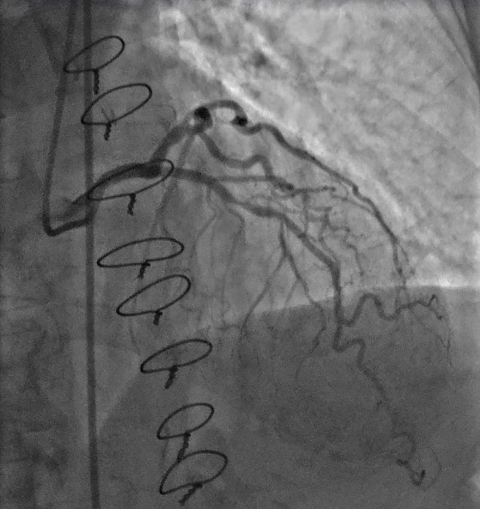
Figure 3. Angiogram showing mid LAD lesion with 60-70% stenosis.
Figure 4.
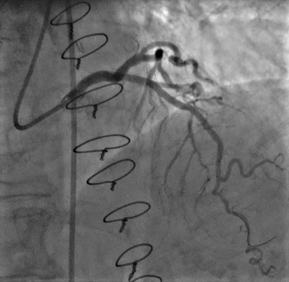
Figure 4. Angiogram after placement of drug-eluting stents showing no residual stenosis.