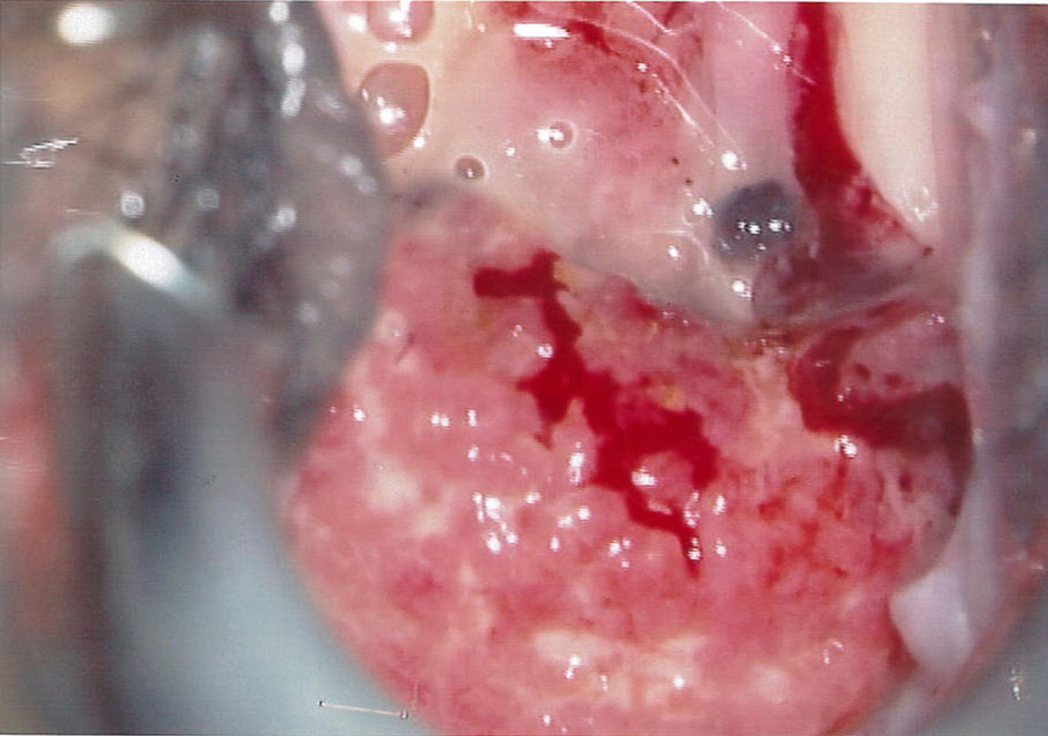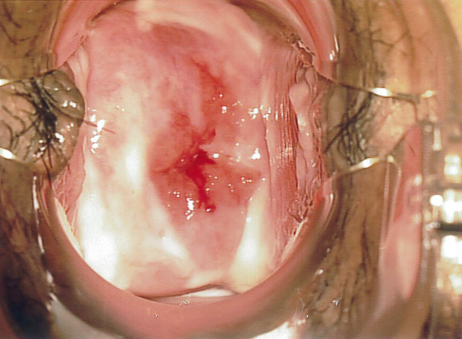
Figure 1. A 3-cm exophytic lesion with contact bleeding noted on colposcopy examination, highly suspicious of malignancy.
| Journal of Medical Cases, ISSN 1923-4155 print, 1923-4163 online, Open Access |
| Article copyright, the authors; Journal compilation copyright, J Med Cases and Elmer Press Inc |
| Journal website http://www.journalmc.org |
Case Report
Volume 9, Number 7, July 2018, pages 204-206
Tuberculosis of the Cervix Mimicking Carcinoma
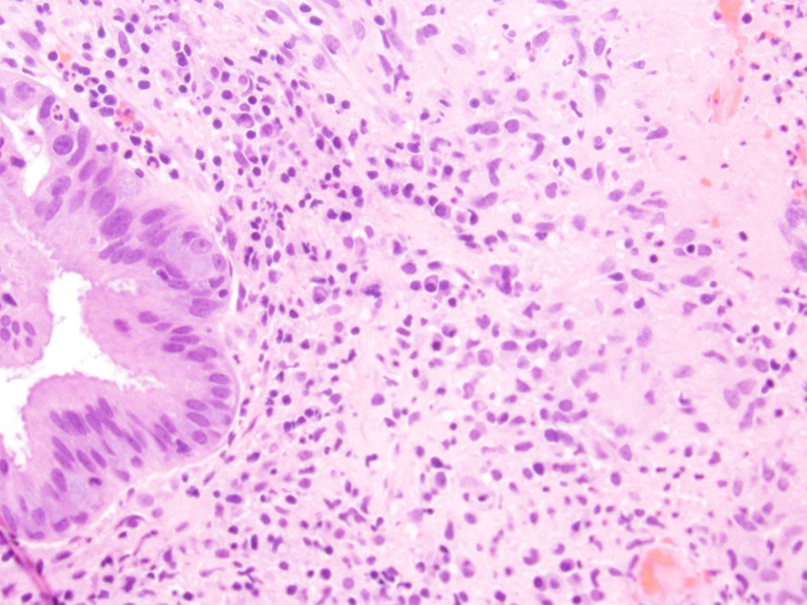
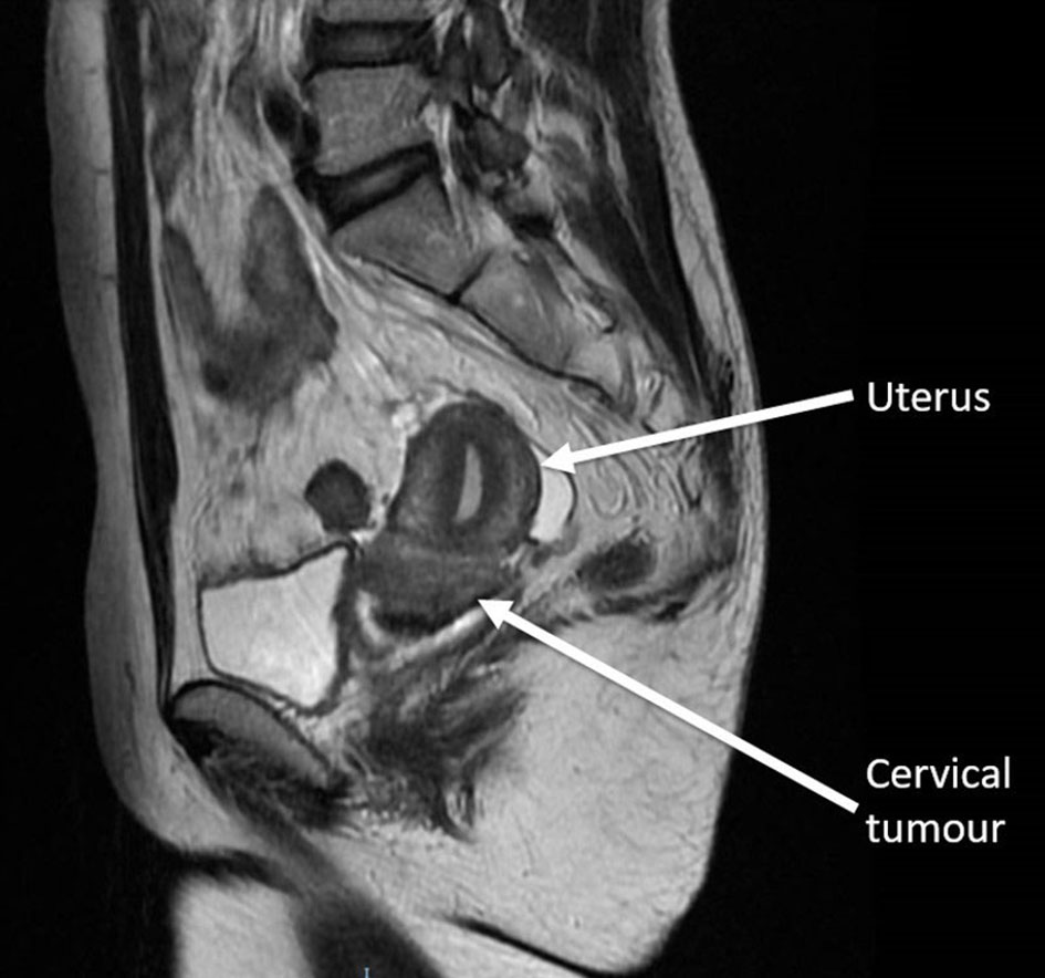
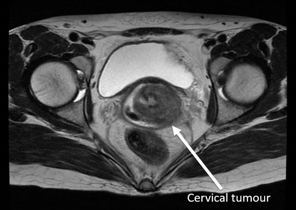
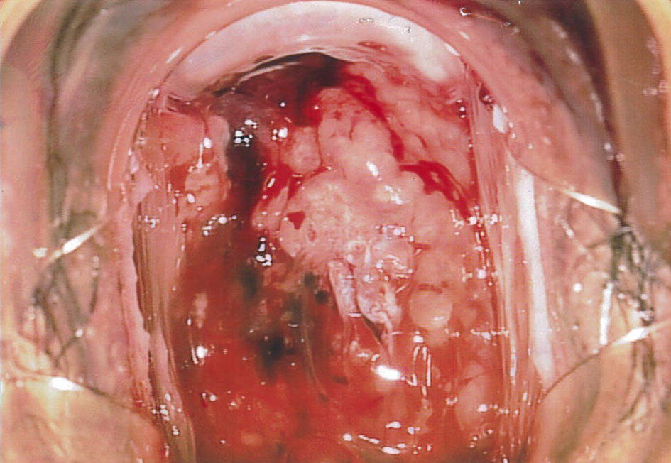
Figures