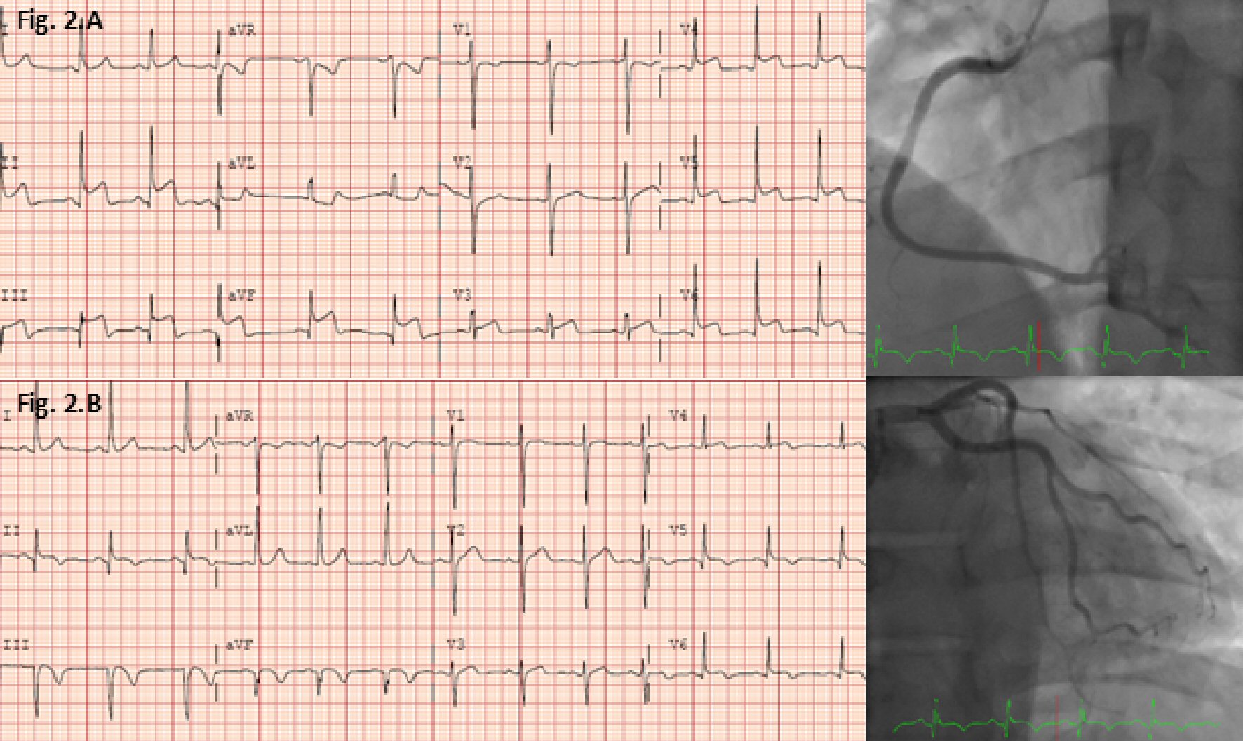
Figure 1. Late gadolinium enhancement (LGE) (yellow arrows) cardiovascular magnetic resonance (CMR) images from first admission. (A) Two chamber view showing mid and epicardial LGE along the inferior wall, and 4 chamber view showing epicardial LGE along the mid inferoseptal wall. (B) Short axis views (basal to apical) showing mid and epicardial LGE along the inferior wall.
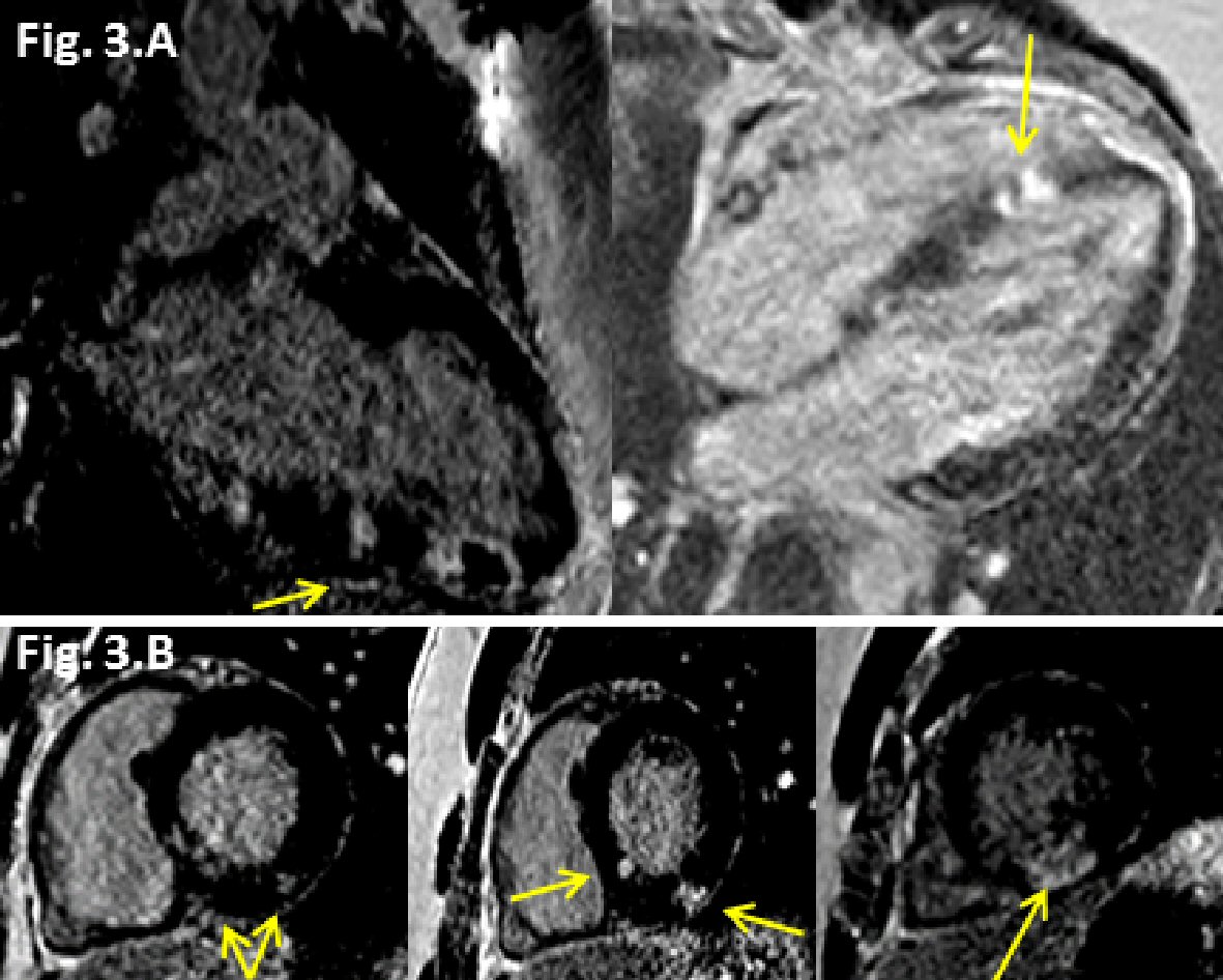
Figure 3. Late gadolinium enhancement (LGE) (yellow arrows) cardiovascular magnetic resonance (CMR) images from the second admission. (A) Two chamber view showing progression of LGE (transmural) along the inferior wall, and 4 chamber view showing progression of LGE (transmural) along the mid inferoseptal wall. (B) Short axis views (basal to apical) showing new patchy epicardial to mid LGE in inferolateral and inferoseptal region walls.