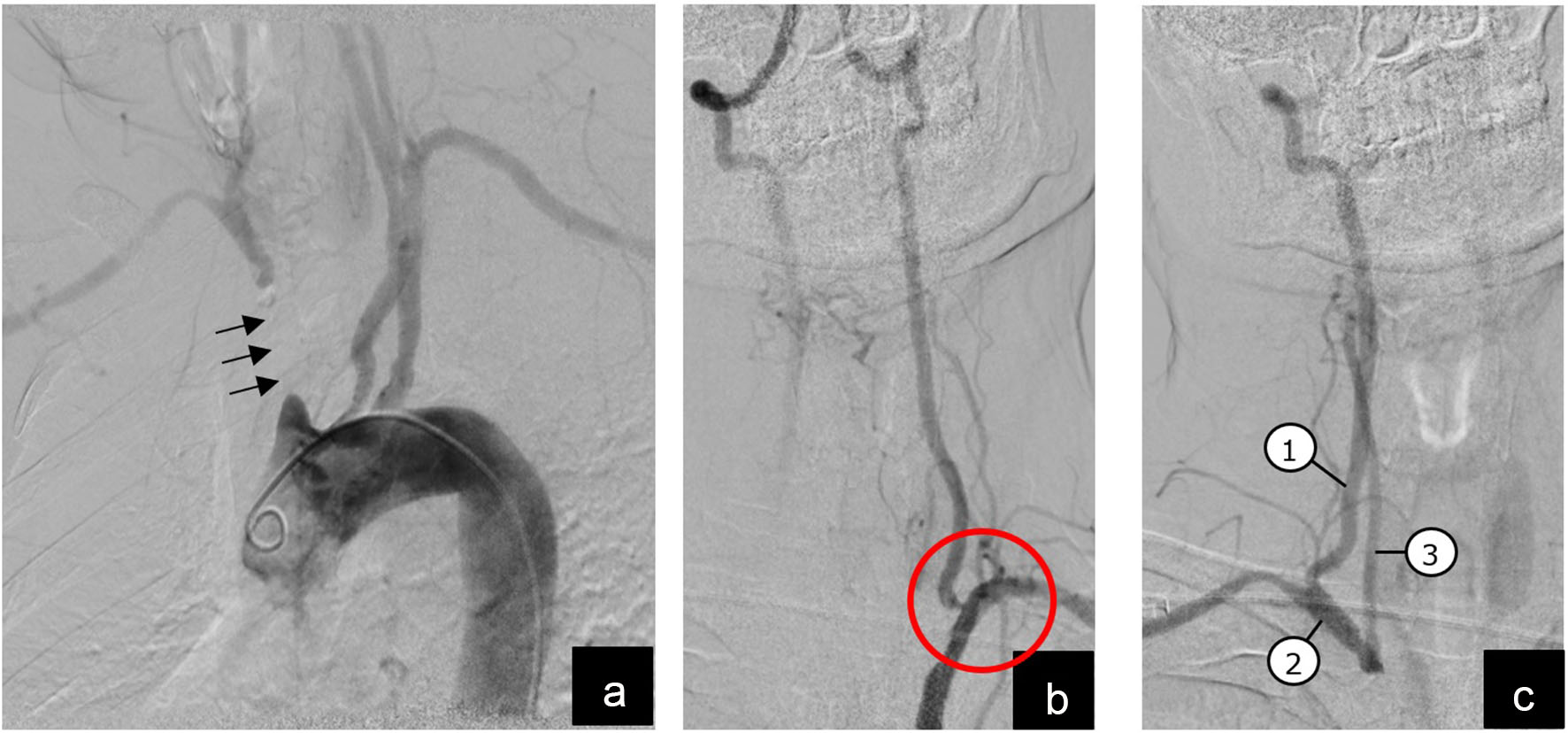
Figure 1. Diagnostic angiography with digital subtraction imaging. (a) Arch aortogram showing blocked segment of innominate artery (arrows); (b) magnified view of left vertebral artery arising from left subclavian artery with a severe stenosis at vertebral origin (red circle); (c) magnified view of right vertebral artery (1) that has a retrograde/caudal flow and fills right subclavian (2) and right common carotid (3) arteries.
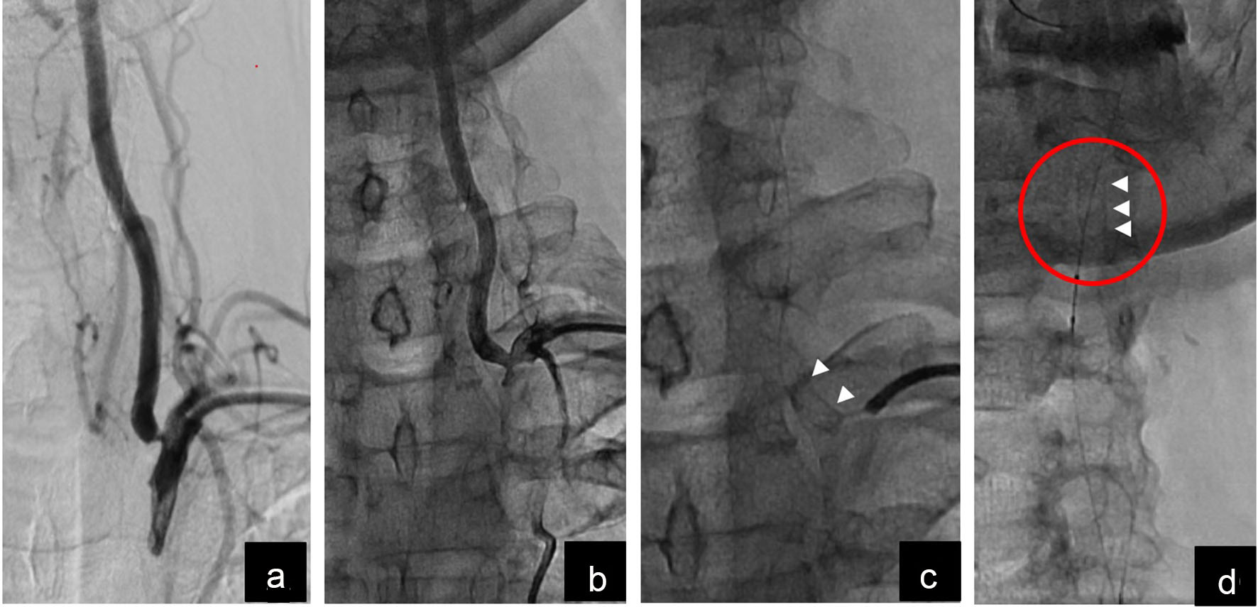
Figure 2. Magnified pictures of left vertebral artery ostial intervention and stent embolization. (a) Vertebral ostium before intervention; (b) partially improved stenosis after ballooning and stenting; (c) short stent visible at vertebral ostium (white arrow heads); (d) embolized stent visualized in distal left vertebral artery above the mandibular line (white arrowheads).
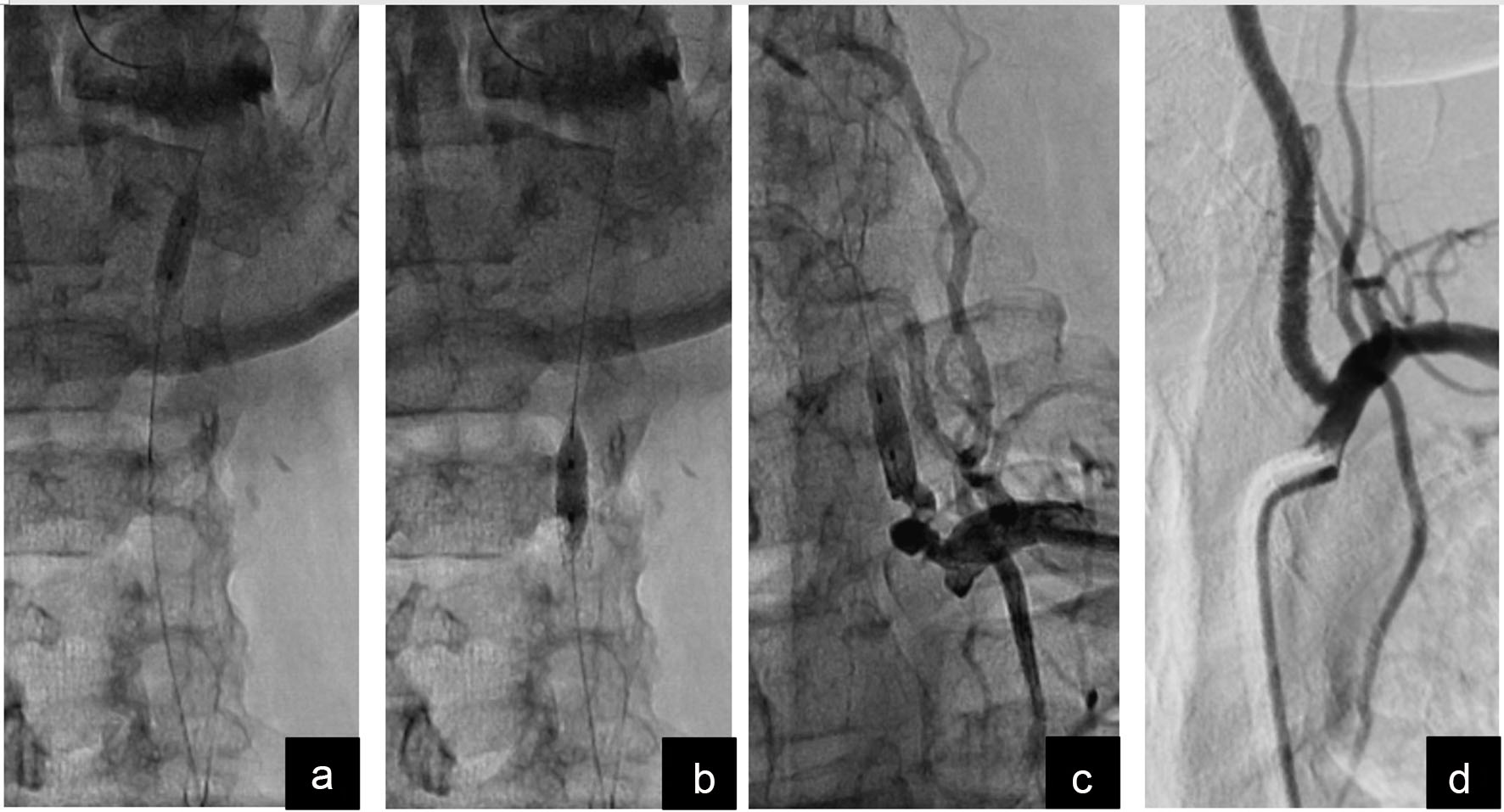
Figure 3. Drag-back technique to manage the embolized stent. (a) Balloon tracked beyond the stent and inflated at low pressure; (b) embolized stent being dragged back by gentle traction on the balloon; (c) embolized stent successfully dragged back to the proximal-most segment of left vertebral artery and anchored against the vessel wall by inflating the same balloon at higher pressure; (d) final result after deploying another stent starting from vertebral artery ostium and overlapping the first stent.