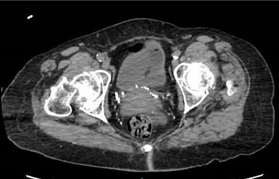
Figure 1. Imaging CT hip: right femoral neck fracture with nondisplaced fracture of the left sacral ala.
| Journal of Medical Cases, ISSN 1923-4155 print, 1923-4163 online, Open Access |
| Article copyright, the authors; Journal compilation copyright, J Med Cases and Elmer Press Inc |
| Journal website http://www.journalmc.org |
Case Report
Volume 9, Number 9, September 2018, pages 309-312
A Collision Tumor With Features of Breast Cancer and Plasma Cell Myeloma as Primary Tumors
Figures
Tables
| CBC: complete blood count; MCV: mean corpuscular volume; RDW: red blood cell distribution; AKP: alkaline phosphatase; ALT: alanine amino transferase; AST: aspartate aminotransferase; ALB: albumin; PT: prothrombin time; APTT: activated partial thromboplastin time; INR: international normalized ratio; LFT: liver function test; BMP: basic metabolic panel; CEA: carcinoembryonic antigen; L: low; H: high. | |
| CBC | |
| White blood cell count | 6.3 |
| Neutrophils | 66 |
| Lymphocytes | 21 |
| Monocytes | 12 (H) |
| Red blood cell count | 3.09 (L) |
| Hemoglobin | 8.1 (L) |
| Hematocrit | 25.6 (L) |
| MCV | 83.0 |
| RDW | 19.3 (H) |
| Platelets | 187 |
| LFT and coagulation factors | |
| AKP | 68 |
| ALT | 10 |
| AST | 53 (H) |
| Bilirubin | 0.5 |
| ALB | 2.5 (L) |
| Protein | 9.8 (H) |
| Globulin | 7.3 (H) |
| PT | 15.5 (H) |
| APTT | 28.8 |
| INR | 1.23 (H) |
| BMP | |
| Glucose | 100 (H) |
| BUN | 20 |
| Creatinine | 0.65 |
| Sodium | 130 (L) |
| Chloride | 100 |
| Potassium | 4.4 |
| Calcium | 9.3 |
| CO2 | 22 |
| Tumor marker | |
| CEA | < 1 |
| SPEP: serum protein electrophoresis; IgA: immunoglobulin A; IgG: immunoglobulin G; IgM: immunoglobulin M; Alpha 1: alpha-1 globulin; Alpha 2: alpha-2 globulin; Beta: beta globulin; Gamma: gamma globulin. | |
| SPEP | |
| Total protein | 10.3 (H) |
| Albumin | 3.2 |
| Alpha 1 | 0.5 (H) |
| Alpha 2 | 0.9 |
| Beta | 0.9 |
| Gamma | 4.7 |
| B2 microglobulin | 3.5 |
| Free light chain | |
| Kappa | 698.0 (H) |
| Lambda | 0.29 (L) |
| K/L ratio | 2,406.90 |
| Immunofixation | |
| IgA | < 19 |
| IgG | 5,300 (H) |
| IgM | < 8 |