Journal of Medical Cases, ISSN 1923-4155 print, 1923-4163 online, Open Access
Article copyright, the authors; Journal compilation copyright, J Med Cases and Elmer Press Inc
Journal website http://www.journalmc.org

Case Report

Volume 10, Number 1, January 2019, pages 8-13


Metachronous and Synchronous Multiple Primary Carcinomas in an Elderly

Figures

Figure 1.
Figure 1. (a) Tonsillar fossa lesion biopsy with high power magnification showing a moderately-differentiated squamous cell carcinoma (SCC) of the left tonsil. (b) Epiglottis biopsy with low and high power magnification showing a moderately-differentiated SCC of the right epiglottis.
Figure 2.
Figure 2. (a) Tongue biopsy with low and high power magnification showing violation of the basement membrane by scattered tongues of malignant epithelial cells. (b) Glossectomy microscopic examination including and revealing SCC.
Figure 3.
Figure 3. (a) Right lung endobronchial biopsy revealing a keratinizing SCC. (b) Chest CT scan showing a 3.5-cm right infrahilar mass that occludes the right lower lobe bronchus.
Figure 4.
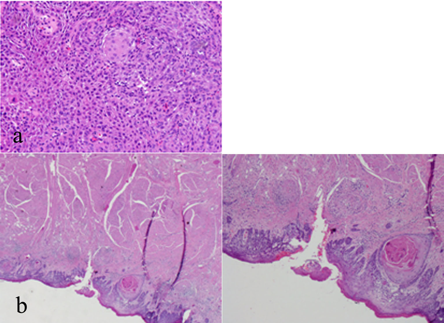
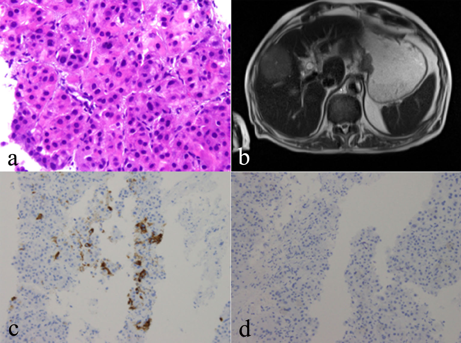
Figure 4. (a) Right liver mass biopsy indicating hepatocellular carcinoma (HCC), biopsy of which shows a trabecular growth pattern composed of thick trabeculae of neoplastic hepatocytes. Smooth contours of this group of HCC cells. Increased nuclear density, plump eosinophilic tumor cells. (b) MRI of the liver revealing T2 hyperintense mass noted in the right lobe of the liver. (c) Hep Par 1 immunohistochemical stain in HCC showing positive cytoplasmic staining. (d) P40 immunohistochemical stain showing no staining, helping to exclude the possibility of metastatic SCC.